A special review published on the 60th anniversary of the discovery of the double helix structure of DNA
Yonat Ashchar and Noam Levitan
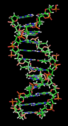
Almost every cell in our body has a nucleus inside which is a well-packaged recipe book containing instructions for building the entire body. If we take the nucleus from one of the cells we can theoretically use it to create a copy of our body, a kind of identical twin or clone, similar to the famous Dolly the sheep. The name of this recipe book, the genome or DNA, is on everyone's lips these days and it is not uncommon to hear or read that "he is a thief, it's in his DNA" or that "humor is in her genes". The ability to read and edit DNA raises hopes, but also fears. People are afraid to eat genetically modified food, into which foreign genes have been inserted, even though every vegetable or animal they consume has - in addition to its own genes - genes from bacteria, viruses and other creatures. Others fear that information collected in their genes, if published, could be used by insurance companies or law enforcement authorities against them. On the other hand, many benefit from the fact that DNA tests make it possible to use this information for the early detection of defects and diseases and to adjust medicines for optimal treatment. Who would have believed that until 60 years ago not everyone recognized that DNA is an important molecule for heredity?
In the first half of the 20th century, almost all scientists thought that proteins, which are much more complex molecules than DNA, are the ones that carry the genetic information. This view changed definitively with the publication of the short article by the American James Watson (Watson) and the British Francis Crick (Crick) in the journal Nature On April 25, 1953.
In the beginning: the discovery of the hereditary material

It can be said that the science of genetics began when Gregor Mendel planted his peas in the courtyard of the monastery, in the 50s of the 19th century. In his hybridization experiments, Mendel showed that the genetic load passes as discrete, indivisible units. The units are organized in pairs and are passed down from generation to generation, with each parent contributing to the offspring of one of the partners. But Mendel's findings said nothing about the material from which the genetic load is made and its location in the cell.
Mendel published his research in 1866 but no one commented on his findings until they were rediscovered in 1900. The famous German zoologist Ernst Haeckel proposed, in the year Mendel's article was published, that the cell nucleus is responsible for the transmission of hereditary traits. Another German, the biologist Walter Flemming (Flemming) showed in 1882 that there is a substance in the cell nucleus that can be colored with a special dye, he called this nuclear substance chromatin (from the word chrome which means "color"). Fleming observed that the chromatin is associated with tiny filamentous structures located in the nucleus and the German anatomist Wilhelm von Waldeyer-Hartz gave them the name chromosomes ("color bodies") in 1888.
Fleming and other scientists discovered how chromosomes organize and behave during cell division. The evolutionary biologist August Weismann (Weismann) was based on their work when he predicted, in 1887, that the gametes must be formed through a special division process that results in cells with half the number of chromosomes in a normal cell. Thus, when a sperm cell and an egg cell unite in the fertilization process, a new cell is created with a normal number of chromosomes. Without this reduction division the amount of chromosomes in the cells would have doubled in each generation.
The preservation of the number of chromosomes during cell division and in the process of fertilization indicated that the chromosomes are responsible for the inheritance of traits, but their connection to the genetic load was clarified only in light of Mendel's findings. Two years after the rediscovery of Mendel's research, the American Walter Sutton noticed, during doctoral studies in zoology (which he did not finish), that chromosomes behave like Mendel's units of heredity. Sutton, who studied grasshoppers, showed in an article published in 1902 that chromosomes come in pairs - one from the father and one from the mother - and suggested that they contain the "genetic factors" that Mendel spoke of. The German biologist Theodor Boveri, who studied sea urchins, noticed that only when the egg and sperm contain all the chromosomes is there normal embryonic development. Bubery claimed, in 1904, that the chromosomes are Mendel's units of heredity and that he reached this conclusion already in 1902. Edmund Beecher Wilson (Wilson), a well-known American zoologist and geneticist, who was Sutton's supervisor and a friend of Bovary gave Sutton's discovery the name Sutton-Bovary theory.
Sutton and Buberry's theory was undoubtedly an important step and it answered the question of the location of the hereditary material, but it did not end the debate about its molecular structure. The chromosomes are large structures, made of many nucleic acids and proteins. Which one is the genetic material itself? It was not yet known.
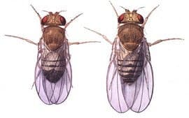
After it was clarified that the genetic load is found in chromosomes, it was Thomas Hunt Morgan (Morgan) who discovered that each chromosome contains genes - referring to Mendel's hereditary units responsible for various traits. Morgan conducted his studies in small flies called Drosophila (Drosophila melanogaster). These flies have been an integral part of genetic research ever since and to this day, so much so that the name given to the species in Hebrew is "Drosophila research". Morgan's research earned him a Nobel Prize in 1933 - the first of many Nobel Prizes awarded in the field of genetics.
In the first half of the 20th century, much progress was made in the study of genes and the form in which they are passed from parent to offspring, but the question of what exactly genes are made of remained open.
The clue to the solution came from an unexpected place. In 1928, the British bacteriologist Frederick Griffith conducted studies on streptococcus bacteria (Streptococcus pneumoniae) that cause pneumonia in humans. These bacteria are often fatal in mice, but some strains are less violent, and do not cause death. Griffith killed the violent bacteria by boiling, then injected them into mice. Perhaps not surprisingly, the dead bacteria did not cause disease. Griffith then mixed the dead, violent bacteria with the live, non-violent bacteria, and injected the mixture into mice. The mice got sick and died. How did a mixture of two substances that separately do not cause disease kill the mice? To answer this, Griffith grew the bacteria that invaded the bodies of the dead mice, and found that they formed colonies similar in shape to the colonies formed by the violent bacteria, not to the colonies formed by the non-violent bacteria injected into the mice.
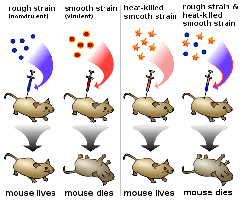
What happened in the bodies of these mice? Apparently, a certain factor moved from the cells of the violent, dead bacteria to the cells of the non-violent bacteria, and changed them - giving them the ability to kill mice, and create colonies in a different way. So, this substance determines the properties of the bacteria - just like the genetic material. Is the "transforming factor", as it was called, actually genetic material?
16 years later, in 1944, three American researchers named Oswald Avery (Avery), Colin MacLeod (MacLeod) and McLean McCarthy (McCarty) decided to check what exactly this transforming factor is. They took the mixture of dead violent bacteria, separated it into its components, and tested each component separately - whether it still had the power to change the properties of the non-violent bacteria. The researchers discovered that sugars, fats and even proteins are unable to do this. Only one component preserved the bacteria's transforming power - deoxyribose nucleic acid, or for short - DNA. This is the same "nuclein" (from the word nucleus, nucleus) a substance rich in phosphorus and lacking sulfur that was first isolated in 1869 by Friedrich Mieshcer, a Swiss physician and biologist, from white blood cell nuclei that he extracted from pus-soaked bandages.
This is how DNA came on the stage of genetics, but not everyone was ready to stand and cheer for the new king. Before the experiment of Avery, McLeod and McCarthy, the conventional wisdom was that the genetic material was a protein. Proteins consist of chains of amino acids, and DNA - of chains of bases (actually nucleotides). There are 20 amino acids that make up the proteins in our bodies, compared to only four DNA bases. To many researchers, it seems much more logical that the complex proteins contain the genetic information - so it can be written in a language of 20 letters, a number smaller than only two of the number of letters in the Hebrew language, for example. How much information can already be stored in a simple molecule like DNA, with only four letters?
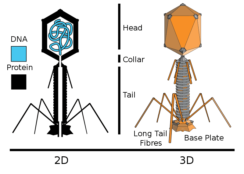
For years the debate between the supporters of the DNA and the supporters of the proteins continued. One of the studies that ultimately led to a decision was a beautiful experiment by two American geneticists, Alfred Hershey (Hershey) and Martha Chase (Chase), in 1952. Hershey and Chase used a system of bacteria and a virus called T2, which attacks bacteria. The virus multiplies by inserting its genetic material into the bacterium, where it multiplies and creates new viruses. To test the nature of the substance that the virus injects into the bacterium, the researchers created viruses with radioactive phosphorus, and other viruses with radioactive sulfur. Phosphorus is found in DNA but not in proteins, and sulfur - only in proteins, not in DNA. They let the viruses infect the bacteria, and after infection, they had bacteria in their hands that contained the genetic material of the viruses. If this material is made of protein, the bacteria infected by the viruses that have the radioactive sulfur, found in proteins, will be radioactive. But if the genetic material is DNA, only the bacteria infected by viruses with radioactive phosphorus, which is in DNA, will become radioactive. As you can guess, the second option turned out to be correct. The viruses labeled with radioactive phosphorus injected the bacteria they infected with radioactive genetic material. The conclusion was clear - the genetic material of viruses is DNA, not protein.
Although the debate continued for several more years, Hershey and Chase's elegant experiment established DNA's position as the king of genetics - a position it holds to this day.
Double helix: the double helix structure of DNA
Even before Hershey and Chase, Avery's experiment convinced Erwin Chargaff, an Austrian Jewish biochemist who left following the rise of the Nazis and immigrated to the United States, of the importance of DNA, and he began researching its composition in 1944. Schragf examined DNA from different organs and biological species and quantified the bases that make it up. The DNA consists, as mentioned, of a chain of nucleotides that differ from each other in the nitrogen base that makes them up and are named after him. The four types of bases in DNA are adenine (A), guanine (G), cytosine (C) and thymine (T). The accepted opinion in Shargaf's time was that all four bases appear in DNA in equal amounts, therefore the molecule was treated not only as simple but also as a boring molecule, repeating itself and unable to contain complex information. In 1950, Sargef discovered something completely different - the amounts of the bases are not equal and the ratio between the bases differs between different species, but not between different organs of the same creature. The difference in DNA composition between different species is another clue that the molecule functions as the genetic material, the recipe book, according to which the organism is built. Another and more important discovery in the matter of deciphering the structure of DNA was that no matter what the total amount of bases, in all the samples he examined the amount of A was always equal to the amount of T and the amount of G was equal to the amount of C. The statement that in DNA there will always be a 1:1 ratio between adenine and guanine to thymine and cytosine is known as "Shargaf's rules". About two years after this discovery, Sargef met with two scientists, who in his opinion were ignorant and arrogant, and explained it to them. These scientists were James Watson and Francis Crick.
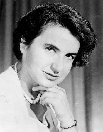
The last campaign in the discovery of the structure of DNA took place in England and was opened at King's College, London, with the joining of the Jewish biophysicist Rosalind Franklin in John Randall's laboratory at the beginning of 1951. In the same laboratory, the physicist Alexander Stokes, the physicist The New Zealander Maurice Wilkins and his research student Raymond Gosling the structure of the DNA molecule using X-ray crystallography, a method in which crystals of the substance being studied are produced and irradiated with X-rays. The rays are scattered as they pass through the crystal, a phenomenon called diffraction, and create A scattering pattern that makes it possible to decipher the arrangement of the atoms in the crystal, i.e. the structure of the studied material. Wilkins was absent from the lab when Franklin, who was an expert in X-ray crystallography, arrived and was told that she would continue Wilkins' research and serve as Gosling's supervisor. The conflict that arose between Wilkins and Franklin was inevitable when he returned from his holiday vacation and discovered that the person he thought was only coming to assist him in his research was a researcher in her own right and was expected to work on his project. In an attempt to reduce the friction between the two scientists, Randall split the DNA study in two, Franklin and Gosling focused on studying a certain form of the DNA crystals and Wilkins and Stokes focused on studying another form whose early results showed that it might have a helical structure.
Wilkins presented at a conference in Naples the photographs of the scattering patterns obtained from the DNA screening. James Watson was also present at the conference, who realized after the lecture that studying the structure of DNA is possible. The 24-year-old American Watson moved to the University of Cambridge to study X-ray crystallography during a postdoctoral fellowship at the Cavendish Laboratory. At the head of the laboratory was Nobel laureate in physics William Lawrence Bragg, one of the fathers of the X-ray crystallography method. Immediately upon his arrival at the laboratory, in October 1951, Watson met Wilkins' good friend Francis Crick, a 35-year-old biophysicist in the stages of finishing his doctorate. The two became good friends thanks to their passion for deciphering the secrets of DNA and, according to Max Perutz, also thanks to their shared intellectual arrogance.
Watson and Crick were brilliant researchers, although not particularly good at conducting experiments, but with an amazing ability to understand, collect and decipher data they received from various sources and, unlike Franklin, were not afraid to make guesses and rely on feelings. Watson traveled in November 1951 to a seminar at King's College and heard a lecture by Franklin about her crystals. At the end of the lecture he quickly returned to Crick and they decided to try to build a physical model of the DNA molecule based on the data he remembered. Building models has proven itself in the past in studying the structure of proteins. Back in the same year, the American biochemist Linus Pauling (Pauling) and his colleagues used this method to discover two structural motifs in proteins - alpha helices and beta surfaces - thereby bypassing Bragg and his team.
The model that Watson and Crick built according to Franklin's lecture was of DNA consisting of three helices with its bases outside. But when Franklin, and the rest of Randall's team commissioned by Watson and Crick, saw the model, she immediately rejected it because it did not match her results. Watson did not understand the data she presented in the lecture at all, apparently because he was busy criticizing her appearance and not listening. Wilkins and Franklin also thought that bothering with building models was a waste of time until all the accurate data was available. Bragg, embarrassed that the people of his laboratory presented a wrong model, forbade Watson and Crick to engage in DNA research and ordered them not to compete with Randall's research but to focus on their protein research.
About a year later, in December 1952, Watson and Crick learned that Pauling and another researcher thought they had found the structure of DNA. Pauling sent a draft of the paper to his son, Peter, in early 1953 at the Cavendish Laboratory and he showed it to Bragg as well as Watson and Crick. To the surprise and joy of the two, the model Pauling presented in the article was wrong and resembled the failed model they themselves built. Even worse, the structure Pauling presented cannot function as an acid and is therefore definitely not the correct structure of the nucleic acid. Watson and Crick realized that Pauling might discover the error and be able to correct the model and that the error would be discovered anyway in February, when his paper was published. When they presented this to Bragg, he agreed that they would go back to working full-force on deciphering the structure of DNA, Pauling had already beaten him in the race to discover the structural motifs of the protein and Bragg would not let him win again!
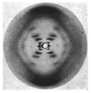
At that time Franklin was already on her way to leave, to her delight, King's College. She was Jewish from a wealthy family at a college largely controlled by the Anglican Church and was looking forward to March when she would leave for Birkbeck at the University of London. As part of the preparations for her departure, Gosling, who was preparing to finish his doctorate without her guidance but under Wilkins' guidance, presented his work to Wilkins. Among the things he presented was a photograph of a DNA scatter pattern that he and Franklin had produced. This excellent photograph, known as "Photo 51", shows an X-shaped pattern that forms when a molecule has a helical structure. Wilkins in turn presented the photograph to Watson when he came to King's College to share with him Pauling's mistake. Watson opened his mouth in astonishment when he saw the photograph and already on his way back to Cambridge drew everything he remembered from it and realized that the structure in question was a double helix. In his book "The Double Helix" he wrote: "Francis will have to agree. Although he is a physicist, he knew that important biological objects come in pairs."
Watson and Crick completed the information they still lacked about Franklin's findings with the help of Max Perutz, Crick's doctoral supervisor who was a member of the Medical Research Council. At the request of the two, Perutz delivered to them a report that Franklin submitted to the council, which contained a summary of the results of her research and various numerical data. Both photograph 51 and the report were shown to Watson and Crick without Franklin's knowledge.
Now, with all of Franklin's data in hand, Wilkins' data and the discovery that Sargef had explained to them about the relationship of the bases in DNA, Watson and Crick combined all the information into one piece and built a model showing DNA as a helix consisting of two strands in opposite directions, head to tail, with the bases facing Inward and arranged in pairs: in front of A will always come T, and in front of G will always appear C. This arrangement also raised a simple possibility for the genetic information replication mechanism, where each strand can serve as a template for the construction of the new strand. If the sequence on one strand is GATTACA, for example, then the sequence on the other strand must be CTAATGT and vice versa. As soon as they connected all the components, on February 28, 1953, they realized that they had deciphered the structure of the hereditary molecule. Crick ran enthusiastically to their favorite pub, "The Eagle" and announced that they had discovered the secret of life. A few days later the model was shown to Wilkins, who confirmed its correctness. Franklin also realized that the model we built was the right one, since it supported much of the data she collected herself and began to organize into articles. If it weren't for Watson and Crick, it is most likely that Franklin would have discovered the structure of DNA on her own within a year or two.
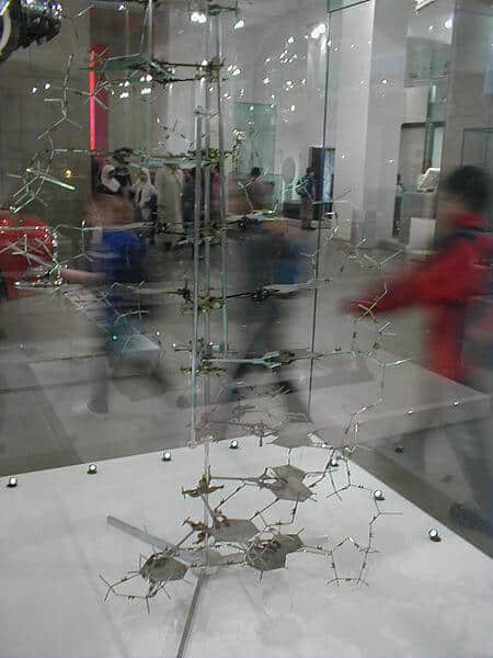
Watson and Crick published the structure they discovered in a short article, a little more than a page long and simply called "DNA Structure", in the journal Nature on April 25. It ends in a British understatement with the sentence: "We have not lost sight of the fact that the particular pairing [of the bases] that we have assumed makes it possible to immediately propose a possible replication mechanism for the genetic material." Along with their article, two more articles were published in the same issue, one by Franklin and Gosling and one by Wilkins and his colleagues, in which the measurements that supported the double helix model are presented. The day the articles are published is the birthday of molecular biology.
Watson, Crick and Wilkins won the Nobel Prize in Physiology or Medicine for their discovery in 1962 (the same year Perutz and John Kendrow also won the Nobel Prize in Chemistry for their discoveries in Bragg's laboratory). Rosalind Franklin died in 1958, at the age of 37, from ovarian cancer. During her illness, she stayed with various friends, including Crick and his wife. The Nobel Prize is only awarded to living people, so Franklin could not accept it. Also, since the award can be divided between no more than three people, it is impossible to know if she would have received it even if she had been alive.
Decoder encryption: Decoding the genetic code
Watson and Crick, as well as Wilkins and other researchers, turned to biological research and an attempt to crack the structure of DNA and the code of life, inspired by the book of the famous physicist Erwin Schrödinger, "What is life?". Schrödinger, who is also known for his abuse of an imaginary cat, claimed in his book that the hereditary molecule must include a type of code according to which the development and activity of the individual is determined. A few weeks after the publication of their first article, at the end of May 1953, Watson and Crick published another article in Nature in which they expanded on the meaning of the structure they discovered for the process of replicating the genetic material and also stated that the sequence of bases in DNA is the genetic code, the cipher, of the hereditary molecule and that mutations can occur following changes in the form or position of the bases.
When Crick and Watson received the Nobel Prize in Chemistry in 1962, Crick dedicated his lecture to the subject that was at the center of genetics science in those years: deciphering the genetic code.
In those years, it was already clear to everyone that DNA is indeed the genetic material. It was also known that certain DNA sequences - genes - translate into the sequence of amino acids that build the proteins. The translation process itself became clearer - Watson dedicated his prize lecture to this process and the role of RNA and ribosomes. The RNA molecule is a nucleic acid that transfers genetic information from the DNA for translation in the protein factories - the ribosomes. The next step is to discover the code: DNA consists, as mentioned, of four building blocks (the bases of DNA), and proteins - of 20 amino acids. How does a sequence of 4 letters translate into a sequence of 20 other letters?
Of course, the translation cannot be one to one - for example, the base guanine codes for the amino acid methionine, and so on. This way we can code for only four amino acids, and we will be missing 16. For the same reason, the genetic code cannot be a sequence of two letters. In such a code, the sequence guanine-thymine, for example, would code for the amino acid alanine. But four letters create only 16 sequences of two letters (four to the power of two). We still lack a code for four more amino acids.
So what about a three letter sequence? Four letters can create 64 sequences of three letters (four to the power of three) - thus providing more than enough code sequences for 20 amino acids.
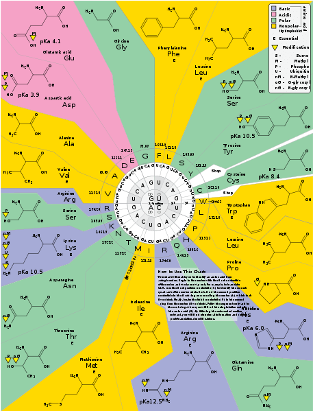
More than enough - maybe too much. To many scientists a code that allows for 44 redundant sequences seems horribly wasteful. What happens when a mutation causes DNA to display a sequence that doesn't code for anything? Did the protein just stop in the middle? Or maybe the amino acids, or at least some of them, are encoded by more than one sequence?
Francis Crick and Sidney Brenner studied the structure of the genetic code by using insertion or deletion mutations - in which there is the addition of one letter to the sequence, or the deletion of one letter. Such a mutation causes the reading frame to shift (Frameshift) and thus disrupts the entire message.
To clarify why such mutations are so harmful, think of a sentence in Hebrew consisting of words with three letters, for example:
One child went to kindergarten alone
If the reading is always in sequences of three letters, if we insert an unnecessary letter, for example another A before the A of "Ehad", we will get:
child aah dahl dog skip n
The entire message has changed beyond recognition.
But, if we cause three insertion mutations, that is, we insert three letters into the sentence, for example, the letters S in the following sentence:
A young boy ran off to kindergarten alone
The result will be three deviations of the reading frame, and in the end - a return to the original reading frame, and the original end of the sentence. Of course, the result will be the same if we remove three letters from the sentence.
Similarly, a gene will produce a normal, or almost normal, protein if it has three insertion mutations or three deletion mutations - but if it has one or two mutations of the same type, the protein produced will be completely different from the original, and often much shorter.
Based on these experiments, Crick and Brenner concluded that the code is indeed made up of sequences of three letters, which were called "codons". Each codon translates to a specific amino acid. For example, the first code that was deciphered was a sequence of three thymines - TTT - which translates to the amino acid phenylalanine.
Now that we know how the code is structured, all that's left is to decipher it.
The breakthrough came following a series of experiments conducted by the biochemists Marshall Nirenberg and Heinrich Matthaei. Nuremberg and Matai used the RNA molecule, the molecule that mediates as mentioned between DNA and the production of proteins. RNA is a nucleic acid with a structure very similar to the DNA molecule, but with uracil (U) instead of thymine. The RNA creates a copy of a certain region in DNA - known as a gene - and this copy is translated into a protein.
Today, it is possible to synthesize a nucleic acid with any desired sequence, and biotechnology companies supply laboratories with DNA sequences upon order. This technology was not available in the early 60s. Nürnberg and Mayai produced RNA molecules simply by creating a solution in which there were only uracil bases. The only sequence that could be created this way is, of course, a sequence consisting entirely of uracil: "UUUUUUUUU...". The protein formed from this sequence consisted entirely of one amino acid - phenylalanine. The first code is decoded - a sequence of three uracil (or three thymine in the DNA molecule) encodes phenylalanine. Similarly Matai discovered that the sequence CCC codes for proline.
The next step was to create a solution containing almost only uracil, but also some adenine. The sequences that can form in such a solution, besides UUU, are UUA, AUU and UAU. And indeed, the protein formed from this RNA was mostly phenylalanine, but it also had leucine, isoleucine and tyrosine. From these results we cannot know for sure which sequence codes for which amino acid - but at least we narrowed down the possibilities...
By creating solutions with different concentrations of different RNA bases, Nürnberg and Matai and other researchers succeeded, until 1966, in deciphering the entire genetic code. In the process, the mystery surrounding the "redundant codons" was also dispersed - it turned out that almost every amino acid is coded by more than one codon, and some are even coded by four.
The discovery that DNA is the genetic material, its structure and how to read the information contained in it opened the way for accelerated developments in the field of biology, thanks to which today it is possible not only to read and understand but also to edit the book of life.
For more information
The original articles [PDF]:
Watson, JD & Crick, FHC A Structure for Deoxyribose Nucleic Acid. Nature 171, 737-738 (1953)
Wilkins, MHF, Stokes, AR & Wilson, HR Molecular Structure of Deoxypentose Nucleic Acids. Nature 171, 738-740 (1953)
Franklin R. & Gosling RG Molecular Configuration in Sodium Thymonucleate. Nature 171, 740-741 (1953)
Avery, OT, MacLeod, CM & McCarty, M . Studies on the chemical nature of the substance inducing transformation of Pneumococcal types. J. Exp. Med. 79, 137-159 (1944).
DNA production at home: According to the Davidson Institute And according to University of Utah.
Originally posted on the blog SciPhile. An article based on this record was published in Galileo Magazine, June 2013
