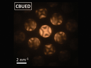
Methods recently developed by researchers from the California Institute of Technology (Caltech) - which make it possible to predict rapid and momentary nanometric changes in the structure of a material in real time

Methods recently developed by researchers from the California Institute of Technology (Caltech) - which make it possible to predict rapid and instantaneous nanometric changes in the structure of a material in real time - were used to simulate the changing electric fields created by the interactions between electrons and photons, and to track changes in atomic structures.
Articles describing the innovative methods appear in the December seventeenth issue of the scientific journal Nature and the October thirtieth issue of the scientific journal Science.
Four-dimensional (4D) microscopy – the approach on which the innovative method is based – was developed at Caltech's Center for Physical Biology for Technology and Fastest Science. This center is headed by Ahmed Zewail, professor of chemistry at Caltech, winner of the 1999 Nobel Prize in Chemistry.
Zewail won the Nobel Prize for pioneering the scientific field of femtochemistry, the use of extremely short laser flashes to observe basic chemical reactions occurring on a femtosecond time scale (quadriillionth of a second, thousandth of a nanosecond). The research "succeeded in capturing atoms and particles during their movement," says Zewail, but while these "photographs" of the particles provide the time dimension of chemical reactions, they do not contribute to the spatial dimension of these reactions - that is, their structure or organization in space.
The researchers were able to observe the missing spatial organization using four-dimensional microscopy, which uses separate electrons to bring the time dimension into typical high-resolution electron microscopy and thus provide a way to observe the changing structure of complex systems at the atomic level.
In the study, described in their article, the scientists were able to focus a beam of electrons into a defined nanometer complex found in the sample, thus enabling them to distinguish point-like nanometer structures at the atomic level. In the method of electron diffraction, a certain object is illuminated by a beam of electrons. The electrons are returned from the atoms of the bone, scatter outward and reach the detector. The patterns obtained in the detector provide useful information regarding the exact spatial location of the atoms in the material being tested. However, if the atoms are in motion, the resulting patterns are blurred, thereby obscuring details regarding minute-scale changes occurring in the material.
The new method developed by the scientists solves the blurring problem by using electron pulses instead of their constant current. The sample being tested - in one case a piece of crystalline zinc - is first heated using a short pulse of laser light. After that, the sample is irradiated by electron pulses at femtosecond time intervals, the electrons are bounced out of the atoms and at the end of the process create the diffraction pattern obtained in the detector.
Since the electron pulses are very short in time, the heated atoms do not have enough time to move much; This short "exposure duration" is what gives the images their sharpness. By adjusting the delay time between the moment the sample is heated and the moment it is photographed, the scientists are able to collect a library of "still" photographs that are then joined together to create a video, similar to the traditional animation process.
"Basically, all the samples we work with are uniform," explains the lead researcher, with very small changes in composition within tiny complexes. "This method provides tools for examining local complexes in materials and biological structures, with a spatial resolution of a nanometer or less and with a time resolution of femtoseconds."
The innovative method of probability enables mapping of the structure of the materials at the atomic level. Using the second method - detailed in an article published in Nature - the light emitted from these nanostructures can be mapped and visualized. The idea behind this method is based on the interactions between electrons and photons. Photons produce temporary fields in nanostructures, and electrons are able to utilize the energy of these fields - which can be observed in four-dimensional microscopy.
In what is known as the "photon-induced near-field electron effect", certain materials - after being hit by laser beams - continue to "glow" for a short, but measurable time (on the order of tens to hundreds femme fatales). In their experiment, the researchers illuminated carbon nanotubes and silver nanowires with short pulses of a laser beam that resulted from previously ejected electrons. The temporary fields persisted for several femtoseconds, and the electrons "collected" energy from them during this time in discrete amounts according to the wavelength of the laser beam.
The advantage of this method lies in the fact that it provides a way to predict the temporary fields when the electrons that received excess energy from them are selectively identified, and that the nanostructures themselves can be simulated with it.
"As described by the reviewers of these articles, this viewing method opens up new realms of imaging possibilities with the ability to influence research fields such as plasmonics, photonics and related fields," notes the lead researcher. "What is fascinating, from the point of view of basic physics, is the ability to simulate photons using electrons. Normally, due to the mismatch between the energy and momentum of electrons and photons, we did not observe the strength of the effect tested with our innovative method or the ability to visualize it in the dimension of time and space together."

9 תגובות
1. Correction to the article: a femtosecond is a millionth of a nanosecond and not a thousandth of a nanosecond.
1. Correction to the article: A femtosecond is a millionth of a nanosecond and not a thousandth of a nanosecond.
2. Instead of calling it four-dimensional microscopy, they couldn't just say - we found a way to photograph atoms on video.
to anonymous,
No one thinks for a moment that this is a non-Arabic name. what are you worried about
and the devil's advocate,
I didn't say no, but if you say..
You can write "Ahmed Zawil". won't kill you (literally)
Eyal
In fact, there is also a probability that the electron will be inside the nucleus and not just "around".
lol! Is that what all the effort is for?
I see photons all the time without a microscope and without plaster.
What is really going on there in the picture?
And Raan, it is true that this is what was taught in school, but it is not accurate. The position of the electrons around the nucleus of the atom can be described probabilistically, so that for every point you choose around the nucleus there will be such and such a probability of the presence of an electron. If you want to expand your knowledge on the subject, search for "orbitals".
Also specifically to your question: http://www.weizmann.ac.il/zemed/net_activities.php?cat=1448&incat=1412&article_id=2097&act=forumPrint
It could be the entanglement of photons
What is this X inside the atom?
Shouldn't an atom look like a nucleus with an elliptical orbit of electrons around it?