Scientists are copying nature's most complex organ, hoping to crack the mysteries of brain diseases, from autism to Alzheimer's
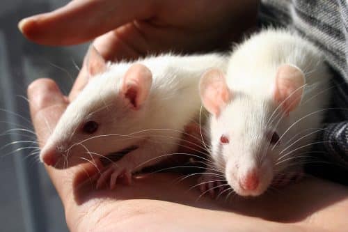
- Knowledge about the human brain is often obtained from experiments on mice, rats, and other animals. The brains of these species are similar to the human brain in many ways, but they lack a highly convoluted surface area, a difference that affects neural function.
- Unique properties of the human brain may explain why studies in rodents have not led to the development of new treatments for brain diseases ranging from schizophrenia to Alzheimer's. This failure encouraged the search for new ways to conduct neuroscience experiments.
- One option is to grow the largest part of the developing brain in laboratory dishes. It is likely that these "organoids" will provide brain researchers with information that could not be obtained from experiments on mice. And they are already being used to study the Zika virus.
"What I cannot build, I cannot understand" - Richard Feynman, 1988
Everything that makes us human is contained in the 1.4 kilograms of yellowish tissue that makes up the human brain. This is where our thoughts develop, this is where we feel love or hate, and this is where humanity's most creative or most evil ideas are born. This nut-like structure is also the most complex organ created by nature. The brain contains approximately 86 billion neurons, or nerve cells, that need to form at the right time, migrate to the right place, and be wired in the right way for us to survive and thrive.
The greatest challenge of modern biology is to understand how the human brain develops and works. Most of what we have learned about this organ since the birth of neuroscience more than 100 years ago has come from experiments conducted on animals, often mice and rats. Scientists could justify this approach because mice and humans share a common brain architecture: they have many identical cell types, and they essentially rely on the same parts of the brain to perform similar mental processes. But humans and rodents differ in one important way: the surface of the mouse brain is smooth, while the human brain is folded into many folds.
This difference can seem insignificant to most of us. But neurobiologists believe that the folds are extremely important to the activity of the human brain. They make it possible to insert many more nerve cells in the same volume, and are also a prominent sign of "intelligent" animals, such as monkeys, cats, dogs and whales. Evolutionary biologists have shown that the folds arise from another difference between mice and humans: nerve cells in many areas of the brain originate from a particular series of precursor cells found only in tiny numbers in mice.
These differences may explain why common genetic mutations that cause severe neurological diseases in humans have almost no effect when they are inserted into the DNA of mice in an attempt to study the mechanisms of human diseases. If the mutations affect the development or maintenance of the normal architecture of the human brain or the function of cell types that are common only in humans, then such studies are doomed to failure. In fact, the unique characteristics of the human brain are probably one of the reasons why studies in rodents have not led to the development of effective drugs for brain diseases such as schizophrenia, epilepsy and autism.
The differences between mouse and human brains have sparked a race of neurobiology experiments to gain more information. Recently developed my laboratory An exciting approach: growing the largest part of the developing brain on a scaled-down scale in a culture dish. These brain structures, called organoids, provide neurobiologists with a model of the human brain that should provide information that cannot be obtained from experiments with mice. Researchers can examine what happens when the plate brain, or mini-brain, is exposed to some pathogen, such as Zika virus, for example, which can damage brain development in the fetuses of infected women, or what happens when an organoid is built through genetic engineering to simulate a brain damaged as a result of a neurological disease.
brain in a saucer (more or less)
My lab started studying organoids in 2012, whenMadeline A. Lancaster, who was then a postdoctoral student in my research group, developed a way to recreate in a culture dish the essential process leading to brain formation in a human embryo during the initial ten weeks of development [See diagram]. The process we developed relies on human cells called stem cells, which have an amazing property called Pluripotency. Pluripotent stem cells are the type of cells found in embryos in early stages of development. When grown in culture under the right conditions, they can produce any type of tissue: nerve, muscle, blood, bone, or any other type. In the embryo, these new cells retain their pluripotency for only a few days. But under laboratory conditions, researchers can keep them in this state permanently and eventually turn them into almost any type of cell they desire.
First, we grow the cells in a liquid that contains all the nutrients needed to grow Neuroectoderm, the part of the embryo that forms the nervous system. When the cells gather into the so-called ball Fetal body, we place the ball in an amazing substance called Matrigel. This gel, similar to the membrane on which the cells rest in the embryo, is produced by cells taken from a cancerous tumor of cartilage in a mouse and grown in a culture dish. Matrigel is rich in factors that encourage cells to divide and prevent them from dying. It serves as a template that is stiff enough for the cells to cling to, but also flexible enough so that its shape can change in response to the cells.
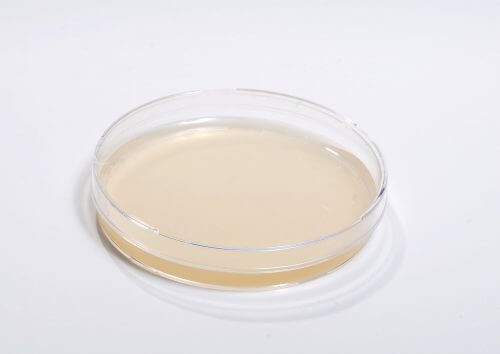
The results of these experiments were truly amazing. When they placed the embryonic bodies on their own in Matrigel, they grew into white, three-dimensional balls of tissue that resembled an embryonic human brain. When stem cells are exposed to the proper chemical signals that encourage brain development in the fetus, they grow and produce perfect replicas of the human forebrain, the area of the brain responsible for our higher mental functions. This area includes ingredients like Cortex (the large outer structure and full of folds) fblood plexus (the area that produces the cerebrospinal fluid). We also saw other structures that guide cells to their proper place in the developing brain. These structures, called the ganglionic eminences the secondary and lateral ones, help in the production of cells that generally moderate the neural activity (interneurons) and help in the production of theHippocampus, involved in creating memories.
Cells in the growing organoid arrange themselves in the same way as those in the brain of an 8-10 week old human fetus. In rare cases, organoids even develop small optic cups, i.e. depressions in the tissue containing colored substances (pigments), as happens when the human eye begins to form. Also, as happens in the developing brain, the cells divide and produce the types of nerve cells found in the fetus. And the neurons send axons: Long extensions that make contact with other nerve cells and build the neural network. Before these networks are formed, the nerve cells migrate from one area to another, similar to what happens in the fetus, and thus make it possible to study what happens when nerve cells end up in the wrong place, as often happens in psychiatric diseases.
on the shoulders of giants
The idea of building tissues in culture is not really new. As with most scientific discoveries, the current wave of organoid research relies on years of research, some of it over a century ago. As early as 1907, the zoologist Henry Wilson showed that some simple animals, such as sponges, for example, can reassemble themselves after being separated into single cells, evidence that the brain is equipped with a plan to connect its many components.
In 1939 he discovered Johannes Holtferter that the different cells in a frog embryo look for each other and renew their form even after they have been completely separated. In the 800s, this finding led to an explosion in the number of "regrouping" studies. In these studies, complex animal organs were created in the laboratory, such as the retina and even the cerebral cortex, by mixing the different types of cells that make them up.
Organoids developed from stem cells have already helped in the study of the Zika epidemic
Based on early regrouping studies conducted in 2006-2010, the late Japanese scientist Yoshiki Sasai Ricken Center for Developmental Biology pioneered the use of pluripotent stem cells to create neural tissue, especially the human retina. In fact, our organoid brain technology is a combination of his methods with the groundbreaking research of Hans Culvers from Utrecht University in the Netherlands, who combined stem cells with Matrigel to develop a culture system that can be used to grow intestinal, stomach and even liver and pancreas tissues.
Apart from drawing conclusions from these early studies, our work makes use of recently developed methods that are revolutionizing the field of biomedical research. One method, known as reprogramming, was developed by a Japanese Nobel laureate Shinya Yamanaka from Kyoto University. Using a simple series of genetic changes, reprogramming turns mature body cells back into pluripotent stem cells, and this can be done in any cell, from skin cells to blood cells. Stem cells from a skin or blood sample can therefore become different types of brain cells, and these cells can be grown into organoids. Because of this, this approach avoids the need to use cells taken from embryos.
Reprogramming makes it possible to grow a suitable organoid from a patient with a genetic disease and compare the result with that of healthy people to look for defects underlying the disease, because a genetic defect in the patient's cells should damage the organoid to the same extent as it damages the developing fetus. In fact, we've already used the organoid method to gain insights microcephaly, a syndrome in which patients are born with an extremely small brain. We discovered that organoids that develop from cells of microcephaly patients are much smaller than normal. Because we can grow patient cells in unlimited numbers, we can now conduct a detailed analysis of the chain of molecular events leading to microcephaly in the developing fetus. This should also be true for other brain diseases: using patients' cells to grow organoids may allow neurobiologists to better understand the problems in brain formation that underlie brain diseases: schizophrenia, epilepsy and other diseases that are difficult or impossible to study in animals.
Organoids derived from reprogrammed cells of healthy individuals may also be effective. Indeed, they have already been useful in the study of the current epidemic caused by the Zika virus, which is hypothesized to have caused microcephaly in some babies born to women who contracted the virus during pregnancy. Many laboratories that work on organoids, first in Brazil and then in the USA, did prove that the virus can cause microcephaly, a connection that would have remained a hypothesis if not for this new method. When organoids are infected with the Zika virus, their nerve cells die and organoids are obtained that are much smaller than those that were not infected, similar to the organoids obtained from our genetic microcephaly patient.
It is likely that organoids will help in the study of the disease caused by the Zika virus. By growing a large number of organoids and infecting each of them with a different strain of the virus from different regions of the world, we can try and understand why the virus causes microcephaly in some regions but not others. We could also use the organoids to test why only some people develop microcephaly after exposure to Zika. Organoids may also be used to identify anchor points or receptors that the virus uses to enter cells, and they may also be essential for testing potential Zika drugs before they advance to human clinical trials.
Another technology that promotes the use of organoids is genomic engineering: a collection of methods that allow researchers to change the genetic code of cells. Organoids engineered to contain mutations suspected of causing disease can allow researchers to determine whether the genetic defects actually lead to disease. Eventually, researchers will be able to test whether correcting these mutations will lead to the creation of healthy organoids. If so, the research could lead to new treatments that would eliminate the effect of the mutations.
The technology can also test whether new drugs affect the brain tissue in the desired way, without the need for animal experiments, thus reducing drug development costs. The organoids will also allow scientists to identify unwanted effects on the development of the human brain, thus preventing the use of harmful drugs during pregnancy. If the infamous drug were tested in this way Thalidomide, which harms the development of the brain early during pregnancy and causes additional birth defects, they probably would not have designated it to prevent nausea during pregnancy in the 50s and 60s.
Organoids are becoming a very important tool for evolutionary biologists as well. They can be used to identify genes responsible for the enormous size of the human brain compared to other primates. Comparing the human genome to that of primates has already revealed genes that may be responsible for cognitive abilities, such as language, that are unique to humans. Understanding the modes of action of these genes has so far remained a matter of speculation only. Scientists will now be able to insert genes from monkeys and apes into organoids to determine how they affect brain development. Researchers could also insert genes or entire regions of the genome into a monkey organoid to make it function more like a human.
Should we be afraid?
It is to be assumed that some people would shy away from the idea of growing a human brain in a dish. Movies like The Matrix pop up in my mind that bring up ideas of brains growing in a lab and developing thoughts or even personalities. These concerns have no basis. The probability that a brain grown in a lab will develop thoughts of its own is zero. An organoid is not a "humanoid" in a jar and it will not become one even in the distant future. Any conscious being must be able to process information from the senses to develop an internal mental representation of reality. Organoids are unable to see or hear and have no sensory input. Even if we were to connect them to a camera and a microphone, it would still be necessary to translate the visual and audio information into a form that would be understandable to these brain cells in the dish, and today such a translation poses an insurmountable technical challenge.
Organoids are not functional brains, but lumps of tissue that mimic the molecular and cellular function of the organ at an extraordinary level of detail. They resemble pieces of tissue removed during brain surgery, rather than sentient beings. However, growing organoids does raise ethical and legal questions. All organoids are made from cells taken from people with certain legal rights. Because of this, laboratory research should meet the same legal and ethical conditions as samples taken from patients in any industrialized country. The patients, of course, have to agree to have their cells used for research. The same legal requirements are also true for organoids. But even when the benefits are clearly explained to donors, the idea that their cells will produce brain-like structures is not always comfortable for them.
What next?
The advantages of this cellular method outweigh any possible disadvantages. Brain organoids laid the foundations for conducting medical experiments and toxicity studies in human tissue, without the need for animal experiments. However, the current version has no blood vessels. The lack of blood vessels poses no difficulty in the early stages of organoid development, but over time cells begin to die from the lack of oxygen and nutrients. Theoretically, it is possible to provide organoids with blood vessels, either with the help of new XNUMXD printing methods, or by growing them from stem cells. Blood vessels are known to grow into the brain, and it may be possible to mimic this process in XNUMXD cultures.
Another challenge is to prepare organoids, which, like a real brain, will have front-back, up-down and right-left axes of direction. Unlike a real fetus that has well-defined body axes, organoids do not have abdomen-back and head-tail axes. Because of this, they develop randomly, so that their parts turn in different directions. In the developing brain, complex signaling systems provide the brain with the sense of up versus down, and the same chemicals may eventually do the same for organoids. Modern biotechnological methods can produce tissue cultures that contain the chemicals needed to encourage cell growth during development. These methods may eventually allow the creation of organoids with a forebrain on one side and a hindbrain on the opposite side.
We have already started promoting the search for ways to overcome these barriers. We have reached technical achievements that we could only dream of a few years ago. Organoids are already starting to help and deepen our understanding of diseases and they also help in drug development. The ability to grow parts of the brain and work with living tissue opened a whole new chapter in biological research, by providing tissue cultures much closer to reality, and sometimes even a reasonable alternative to using animals for research.
good to know
The emergence of mini-brains to discover what makes us human: a lecture by researcher Madeline Lancaster, who participated in the studies described in the article:
for further reading
- Organogenesis in a Dish: Modeling Development and Disease Using Organoid Technologies. Madeline A. Lancaster and Juergen A. Knoblich in Science, Vol. 345, page 283; July 18, 2014
- Generation of Cerebral Organoids from Human Pluripotent Stem Cells. Madeline A. Lancaster and Juergen A. Knoblich in Nature Protocols, Vol. 9, pages 2329–2340; October 2014
- Dishing Out Mini-Brains: Current Progress and Future Prospects in Brain Organoid Research. Iva Kelava and Madeline A. Lancaster in Developmental Biology. Published online July 9, 2016
- Grow your own eye, Yoshiki Sasai, Scientific American Israel, February 2013
- Diseases of the plate, Steven S. Hall, Scientific American Israel, June-July 2011

One response
It is assumed that in the future the technological challenge of building and connecting a sensory input means - will be solvable. If an organoid with "personality" is indeed obtained - it will be a scientific achievement that will overshadow every scientific achievement achieved in the past. It will be possible to redefine the model of human reason and build new strengths of human reason. The discussion of the psychophysical problem will also be promoted, although it is highly doubtful that a solution will be reached on this issue.