Weizmann Institute of Science scientists have discovered coil-like cholesterol crystals that form in the cells of the immune system that fight atherosclerosis
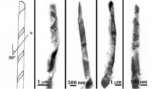
Helix-shaped cholesterol crystals. Digital processing of data obtained from four different crystals in different cells using soft x-ray imaging under cold conditions (cryo-SXT)
No bad cholesterol. Without cholesterol we would not live - as this substance gives the cell membrane the necessary character for its function. Even the LDL particle, which is notorious as a risk factor for the development of vascular diseases, is not "bad" in itself. In order for it to move through the bloodstream and reach the body's cells, cholesterol is packed inside LDL particles that allow it to dissolve in the bloodstream. Cholesterol crystals, on the other hand, are already a bundle of trouble. As long as cholesterol is in its molecular state, the body deals with it - even if it is in excess, and even when it accumulates in the arteries. But when cholesterol crystallizes - the body no longer has a way to deal with it, and the accumulation process becomes irreversible. Although atherosclerosis is the main cause of heart attacks and strokes, one of the leading causes of death in the West, it was not known until now how these crystals are formed. In a study recently published in the Proceedings of the American Academy of Sciences (PNAS), scientists from the Weizmann Institute of Science discovered two ways in which cholesterol crystals are formed inside the cells of the immune system that are sent to fight sclerosis.
Why are sclerotic lesions formed in the arteries in the first place? As is known, when there are high levels of LDL cholesterol in the blood, the probability increases that these particles will settle and enter the artery wall, where they will accumulate and oxidize. Since the oxidized LDL is toxic to the cells of the artery wall, the immune system sends monocytes to the scene, which differentiate into macrophages - those "Pacman" cells that swallow unwanted substances and clean the area. At a certain point, after ingesting a considerable amount of cholesterol, the macrophages themselves may turn into very fat cells, called "foam cells", and even die. In this way, remnants of dead cells and fatty molecules gradually accumulate in the sclerotic lesion, the inflammation worsens - and at some point cholesterol crystals also appear. "The accumulation of cholesterol crystals is an essential step in the pathological chain of events that includes cell death, an increase in the inflammatory response and finally the eruption of the sclerotic lesion into the bloodstream. Apparently, as long as there are no crystals, the situation is reversible," explains Prof. Lea Addi from the Department of Structural Biology. "When examining sclerotic lesions originating from humans and animals, cholesterol crystallization is already at a very advanced stage. Crystals are seen outside the macrophages, but also inside them, and it is difficult at this stage to assess where they were formed and at what developmental stage of the disease they sank," adds Dr. Neta Versano from Prof. Addi's laboratory, who led the research in long-term collaboration with Prof. Leslie Lazerovitz from the department for materials and surfaces, expert in crystallography. "In this research we tried to understand how the crystals are formed in the first place. For this we had to develop a method that would allow us to identify crystals in their initial state."
The research group relied on a model that simulates atherosclerosis by feeding macrophages in a plate with LDL particles. To enable a look at the surface of the macrophages and to unequivocally identify the cholesterol crystals at the beginning of their development, the scientists developed a first-of-its-kind correlative method that combines super-resolution fluorescence microscopy (STORM) with soft X-ray imaging under cold conditions (cryo-SXT). The soft X-rays allow a three-dimensional photograph of the crystal and the biological environment in which it grows, with a resolution of tens of nanometers, while the fluorescence microscope allows the crystals to be identified as cholesterol crystals. In the initial stages, crystals in the form of parallel plates were identified near the cell membrane with a size of 200 nm. These crystals have the same shape as cholesterol monohydrate crystals that sink under laboratory conditions. However, in addition to the small plates, thin and elongated intracellular crystals with a size of tens of microns were also detected. Prof. Addi says: "It was a big surprise. We identified large, unfamiliar crystals impaling the cell. We have never seen cholesterol crystals that look like this under test-tube conditions." The structure of the elongated crystals was determined through electron diffraction under cold conditions with the assistance of Dr. Nadav Elad from the Department of Chemical Research Infrastructures, and was characterized as cholesterol monohydrate with monoclinic symmetry - different from the triclinic one of the plates. The elongated crystals were arranged in the unusual shape of a hollow cylinder or coil, similar to the crystals formed in the pathology of gallstones.
The crystal is the template of his birth landscape
The elongated shape of the crystals in the model matches the shape of the crystals in the body, although the latter have triclinic symmetry. Dr. Versano explains: "We believe that the crystals in the body preserve their shape, even in advanced stages of crystallization, the imprint of the process in which they were created. That's why they probably remain elongated, even if they undergo a structural change to create a triclinic arrangement." These findings may pave the way for cracking the crystallization mechanisms of cholesterol in the body, and later even affect the crystallization in a way that prevents or reduces the process. "Now we will try to play with the model and influence cholesterol crystallization in different ways. At the same time, we will examine samples of sclerotic lesions, and we will examine differences between people in the development of the crystals", Prof. Addi describes the future directions for the research. Also participating in the study were Dr. Tali Dadosh and Dr. Ido Penkas from the Department of Chemical Research Infrastructures at the Institute, Fabio Baggi from the Department of Chemistry at the University of Milan, Dr. Eva Ferreiro and Dr. Ana Perez-Barna from the ALBA Synchrotron in Spain and researchers from Prof. Howard Kroth's group from the unit for NIH Experimental Atherosclerosis.
"A great research challenge"
"Pathological processes of crystal formation are a great research challenge because it is a crystallization that is not controlled by the body; This is exactly what the body wants to prevent," says Prof. Leah Eddy, who dedicates her professional life to the study of crystals in living beings. Recently, Prof. Addi was elected as a member of the American Academy of Sciences, and this is her entry article for the position. "In many ways, this research represents me in all my incarnations and all my thought forms. This work combines organic chemistry, crystallography and the use of innovative imaging techniques at the forefront of science, while collaborating closely with my expert colleagues."

2 תגובות
Hello Mr. Levy
Below is a brief explanation of what "arteriosclerosis" is - by Professor Aviram Michael from the Technion School of Medicine - one of the leading lipid researchers in Israel and the world:
[From the Facebook group of Guy Ben-Zvi - an autodidact with a vast amount of knowledge about metabolism
In the human body and especially in body fats - it is very worthwhile to read carefully and follow the posts in the group]
"... what really oxidizes and what makes oxidized LDL so dangerous is the omega-3 in the LDL... and the role of the antioxidants in the blood (vitamin C, polyphenols, etc.) and in the LDL itself (vitamin E) is to make sure that the omega-3 is not oxidized on its journey to the body's cells."
So this is it friends - oxidized LDL (the one that doctors claim causes heart disease) is oxidized omega-3. Oxidized omega-3 is harmful in two ways - it increases the oxidizing load (peroxides of omega-3 are radicals in themselves) and causes a lack of "fresh" omega-3 in the cells.
And what do we do about it? What I have been saying for 15 years - consume omega 3 and vitamin E.
https://www.facebook.com/groups/886489568048219/permalink/1469821633048340/
Greetings!
very interesting
It is very important for me to know if it is possible to clean arteriosclerosis.
Is the project in progress and if so am I ready to participate in it?
In any case, I suffer from severe sclerosis after 10 catheterizations
and feels very bad and is waiting for a more effective solution.
Sincerely
Avi Levi
Mobile 0543372901