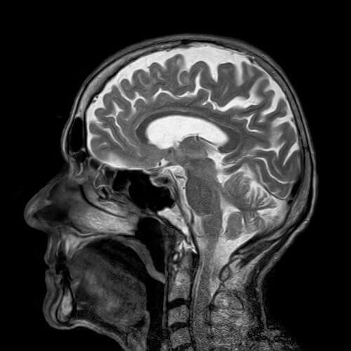
Dr. Anaho Gong, founder of Subtle Medical of Menlo Park, which develops computing in the field of medical imaging, presented a deep computer learning algorithm that was able to display high-quality MRI scan images at a dose of 10% of the amount of gadolinium that is usually given

Several studies done in the last year revealed that gadolinium enters our body, but does not actually leave, but accumulates. It is not yet known whether this has health consequences, but already today research institutes are trying to find methods in order to save the amount of gadolinium. A new study led by Dr. Enhao Gong, founder of a computing company in the field of medical imaging from California in the United States called Subtle Medical of Menlo Park, presented at the last annual conference of the Radiological Society of North America (RSNA) a deep learning algorithm. who was able to show high-quality MRI scan images at a dose of 10% of the amount of gadolinium that is usually given. The researchers also claim that there is no obstacle to using the algorithms to reach results of 5% of the amount of gadolinium that is used today, or even lower than that.
Deep learning is a field of research in the world of computers that aims to computer-imitate the operation of the human brain. What is special about deep learning systems is their ability, similar to the human brain, to learn and improve all the time. Through the use of models called "conventional neural networks", the computer can recognize not only images, but also find subtle differences between the image data, differences that a human observer is unable to distinguish between them. The new algorithm learned to estimate the scans with the full dose of gadolinium from the scans with the low dose of gadolinium.
For the purpose of the study, the researchers used magnetic resonance images from 200 patients who had MRI scans with contrast material for a variety of indications.
The team collected three sets of scan images for each patient: the first set was a set of scan images before the injection of contrast material, which they referred to as scans with zero contrast material. The second group was a group of scan images after the injection of a low amount of gadolinium, scans in which 10% of the amount that is usually injected was injected - these images were processed by a deep learning algorithm. The third group was a group of scans in which the full amount of gadolinium that is usually injected today was injected. For each subject, these three scan images were taken and they were given to neuroradiologists (radiologists who specialize in imaging the brain and spine) to evaluate the effect of the contrast agent and their general quality
The radioneurologists could indeed distinguish which of the scans had a reduced amount of 10% gadolinium, but the information they received from these images following the processing of the algorithm was quite the same as the information they received from the scans with the full amount of gadolinium.
The research is currently moving to clinical stages and evaluation of the algorithm across a wider range of MRI scanners and with different types of contrast agents in order to improve the imaging capability that exists today while saving the contrast material.
- More information about MRI and gadolinium can be found in the book "MRI the complete guide-medicine and physics meetOn the website mriguide.co.il
Link to the research summary - RSNA website
Gadolinium in MRI - is it dangerous for us? - from the Israeli MRI portal

3 תגובות
Indeed, an unnecessary word correction of Word... (thanks to someone).
No external devices and always and daily
Ofer Hi, this is a convolutional neural network and not conventional networks as you wrote.