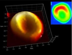
Scientists using an electron microscope and nanometer-sized scanner were able to image a red blood cell and also detected signs of the blood's coagulation material in the wound caused by the arrow that struck Utzi's back

His DNA was deciphered, the intestinal and stomach samples allowed us to reconstruct his last meal. The circumstances of his violent death have already been explained. However, what eluded the scientists was the identification of traces of blood in the body of Otzi, a young man whose body was preserved as a mummified body in a glacier in Switzerland. An examination of his aorta yielded no results. However, recently, a team of scientists from Italy and Germany, using nanotechnology, was able to locate the red blood cells in the wounds on Utzi's body, thus discovering the earliest traces of any human red blood cells.
"Until now, there was no certainty about how long the blood could survive - not to mention the blood cells of humans from the Chalcolithic, Stone and Copper Ages. " Says Albert Zink, head of the Institute of Mummies at the EURAC European Academy in Bolzano, Switzerland. Zink explains the starting point of the research he conducted together with Mark Janko and Robert Stark, materials scientists at the Technical University of Darmstadt. Even for modern forensic medicine it is difficult to almost impossible to determine how long traces of blood can survive at a crime scene. The three are convinced that they succeeded in developing nanotechnological methods in which they tested Otzi's blood and were able to analyze blood microspheres and tiny blood clots that might lead to a breakthrough in this field.
The team of scientists used an atomic force microscope to study thin sections of tissue from the arrow gaping wound that entered Otzi's back and a scratch on his right hand. The device scans the surface and the cavities of the embroideries. Sensors measure every small deviation of the test line, point by point, and build a XNUMXD visualization of the surface. The result was a picture of classic "doughnut-shaped" red blood cells, just like we find in healthy people today.
"To be absolutely sure that we are not seeing remains of pollen, bacteria or even the imprint of a blood cell, but actual blood cells, we used an analytical method known as Raman spectroscopy," reads the report in Raman spectroscopy. identify different molecules. According to the scientists, the images derived from this process match modern-day human blood samples.
While examining a wound at the point where the arrow entered the body, a team of scientists also identified fibrin, a protein involved in blood clotting. "The fibrin is found in fresh wounds and then its quantity deteriorates. According to the theory, Otzi died a few days after being wounded by an arrow, now this theory has been confirmed, Zink explains.
The team published their research in the Journal of the British Royal Society of Sciences.

2 תגובות
At least proofread the title line.
Electrons, put up…