"For decades, the field of electrophysiology has been based on the use of cells and cultures grown on two-dimensional surfaces, an innovative 'organ-on-a-chip' technology," explains Prof. Tzachi Cohen-Karni
[Translation by Dr. Nachmani Moshe]
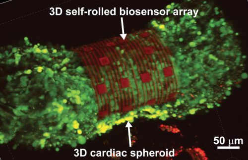
An international team of researchers from Carnegie Mellon University and Nanyang Technological University in Singapore (NTU Singapore) succeeded in developing an organ-on-a-chip system that uses bioelectrical sensors to measure the electrophysiology of cardiac cells in a three-dimensional array
These XNUMXD biosensor arrays, which wrap around elliptical heart cell tissues and wrap them tightly, create an 'organ-on-a-chip', and thus allow researchers to examine how cells communicate with each other in multicellular systems such as the heart.
The 'organ-on-a-chip' approach could help in the development and evaluation of the effectiveness of drugs to treat diseases, and perhaps even allow researchers to screen drugs and toxins directly on a human-like tissue, instead of testing them inside human tissues. The system can also be used to gain insights into the relationship between the electrical signals in the heart and the development of diseases, for example heart rhythm disorders. The research, which has long been published in the journal Science Advances, allows researchers to examine processes within cultured cells that are currently unavailable to researchers, processes such as tissue development and cell maturation.
"For decades, the field of electrophysiology has been based on the use of cells and cultures grown on two-dimensional surfaces, such as Petri dishes," says Tzachi Cohen-Karni, professor of biomedical engineering and materials science at Carnegie Mellon University. "We are trying to face the challenge of measuring the heart's electrical patterns in a XNUMXD image by developing sensors that wrap around the heart cells and thereby provide electrophysiological information from this tissue." The device-on-a-chip system starts out as a small flat rectangle, similar to a diminutive bar bracelet. A bar bracelet starts out as a rigid, rolled structure, but when you release the tension within it, it soon wraps around your wrist. An organ-on-a-chip begins similarly. The researchers fix a system of sensors made of metallic electrodes or sensors of the material graphene on the surface of the chip, then eat away the bottom layer made of germanium, known as the "sacrificial layer". Once this layer is removed, the array of sensors is released and wraps the surface in a structure similar to the shell of a barrel.
The researchers tested the system on organoids composed of heart cells. These three-dimensional structures of heart cells are 3-2 times the width of a human hair. Wrapping the cells allows researchers to collect readings of electrical signals with high precision. "In practice, we created a system of XNUMXD biosensors to measure the electrophysiology of stem cells that then differentiate into heart cells," explains the lead researcher. "This system could be used in the study of the regeneration and development of heart tissue, and potentially be used to treat damaged tissue after a heart attack, for example, or for the development of new drugs to treat the disease."
"Mechanical analysis of the wrapping process allows us to precisely control the shape of the sensors, in order to make sure that there is close contact between them and the heart tissue," says the lead researcher. "The method also makes it possible to automatically adjust the level of tightness of the delicate contact between the sensors and the tissue so that high-quality electrical signals can be measured without changing the physiological conditions of the tissue due to external pressure."
"The general idea is to take methods that are normally performed in planar geometry (two-dimensional) and adapt them to volumetric-spatial geometry (three-dimensional), the researcher explains. "Our organs are three-dimensional by nature. For many years, electrophysiology has been measured using cells grown in cultures on a two-dimensional tissue surface in a Petri dish. However, now, these amazing electrophysiology methods can be applied with the help of three-dimensional structures."
such as Petri dishes," says Tzachi Cohen-Karni, professor of biomedical engineering and materials science at Carnegie Mellon University. "We are trying to face the challenge of measuring the heart's electrical patterns in a XNUMXD image by developing sensors that wrap around the heart cells and thereby provide electrophysiological information from this tissue." The device-on-a-chip system starts out as a small flat rectangle, similar to a diminutive bar bracelet. A bar bracelet starts out as a rigid, rolled structure, but when you release the tension within it, it soon wraps around your wrist. An organ-on-a-chip begins similarly. The researchers fix a system of sensors made of metallic electrodes or sensors of the material graphene on the surface of the chip, then eat away the bottom layer made of germanium, known as the "sacrificial layer". Once this layer is removed, the array of sensors is released and wraps the surface in a structure similar to the shell of a barrel.
The researchers tested the system on organoids composed of heart cells. These three-dimensional structures of heart cells are 3-2 times the width of a human hair. Wrapping the cells allows researchers to collect readings of electrical signals with high precision. "In practice, we created a system of XNUMXD biosensors to measure the electrophysiology of stem cells that then differentiate into heart cells," explains the lead researcher. "This system could be used in the study of the regeneration and development of heart tissue, and potentially be used to treat damaged tissue after a heart attack, for example, or for the development of new drugs to treat the disease."
"Mechanical analysis of the wrapping process allows us to precisely control the shape of the sensors, in order to make sure that there is close contact between them and the heart tissue," says the lead researcher. "The method also makes it possible to automatically adjust the level of tightness of the delicate contact between the sensors and the tissue so that high-quality electrical signals can be measured without changing the physiological conditions of the tissue due to external pressure."
"The general idea is to take methods that are normally performed in planar geometry (two-dimensional) and adapt them to volumetric-spatial geometry (three-dimensional), the researcher explains. "Our organs are three-dimensional by nature. For many years, electrophysiology has been measured using cells grown in cultures on a two-dimensional tissue surface in a Petri dish. However, now, these amazing electrophysiology methods can be applied with the help of three-dimensional structures."
More of the topic in Hayadan:
