Surgeries to sever a lobe of the brain were the solution to epilepsy, but it turned out that we did more harm than good. Now researchers at the Weizmann Institute of Science explain the mechanism that links the two parts of the brain, and why damage to it endangers the patient
By Prof. Ilan Lampel, Yael Oren and Dr. Yonatan Katz, Weizmann Institute of Science

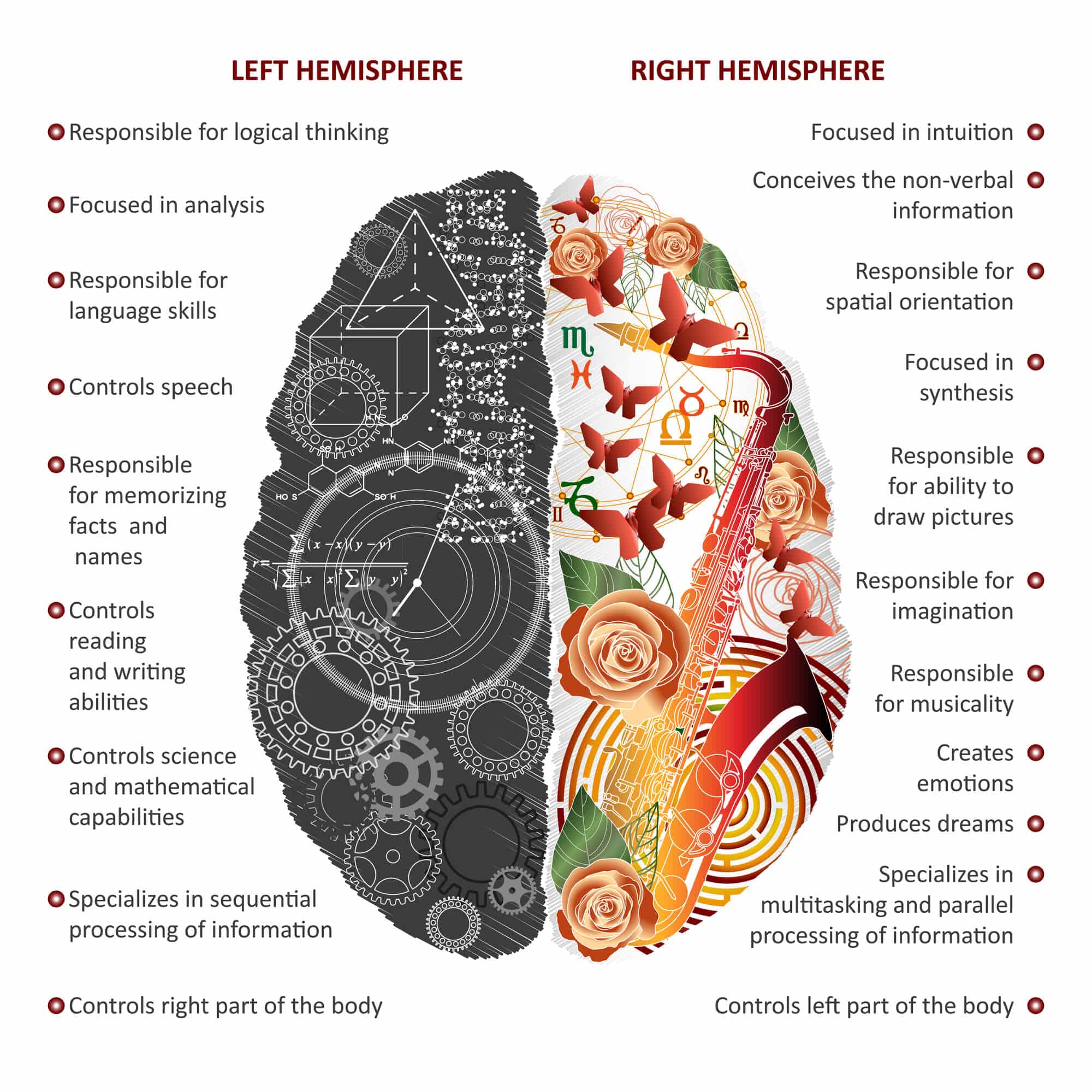
One of the strangest phenomena ever observed in brain research was seen in patients where the two halves of the brain were surgically separated.
split brain
These surgeries were common in the past as a last resort for severe epilepsy patients. In these operations, the surgeons cut the cerebral cortex, the bundle of fibers that connects the two hemispheres. When the separation was almost complete the patients seemed to have two minds controlling one body and sometimes in opposite ways. For example these patients dressed themselves with one hand and at the same time tried to take off their clothes with the other hand. In addition, when they were asked to draw or name an object placed on the right side of their visual field they successfully answered the task, but when the same object was placed on the left side of their visual field they claimed to have seen nothing.
Research in these patients, who were called half-brained, led to the awarding of the Nobel Prize to Roger Sperry in 1981. Although it was known that the half of the brain on one side responds to stimuli on the other side of the body and controls it, Roger Sperry's research led to the understanding that the brain is not completely symmetrical and that The brain plays a central role in transferring information between the two halves of the brain. Additional studies in humans and other mammals showed that the neural activity in corresponding areas in both halves of the brain tends to be very synchronized and in light of many other studies it seemed that the cerebellum plays a central role in this synchronization.
As a basis for starting our research, we thought that if it would be possible to understand how the activity is coordinated between the two cerebral hemispheres, each of which seems to operate as an independent unit, it would be possible to gain insights into how other areas of the cerebral cortex communicate. Therefore, we tested in behaving mice how the behavioral state of the mouse affects the synchronization between the two hemispheres and how this synchronization of information depends on the activity of the cerebral cortex. When we recorded the electrical activity from neurons in somatosensory areas in both hemispheres at the same time, we were surprised to find that the synchronization decreased precisely when the animal demonstrated an active behavior similar to the activity that appears when mice try to gather information from the environment.
In order to examine the role of the cerebellum in the modulation of synchrony in different behavioral states, we blocked the passage of information through the cerebellum for a limited time by inventing genetic tools that combine chemistry ('as genetics'). After the block there was a significant decrease in the high synchrony observed when life was passive, but there was no change in the level of synchronization when life was active. That is, the modulation of the synchronization in different behavioral states was affected by blocking the information that is transmitted to me by the cerebellum. This result hinted at the possibility that the activity of the cerebral cortex decreases while the mice are active and increases during rest. In order to test this, we used a two-photon microscope to monitor the activity of the axons that make up the cerebral cortex while the animal is awake and behaving. The use of this microscope did show that when the mouse was inactive, i.e. at rest, the activity of the corpus callosum was higher compared to times when the mouse was more active, therefore explaining the increase in synchronization during rest. In addition, we found that the average activity of the cells in that area actually decreases, which suggests that the population of cells that connects the two hemispheres is unique.
One of the conclusions from our experiments is therefore that changes in behavior cause a specific modulation on the cells from which the axons that make up the cerebral cortex come out so that their activity level is opposite to the activity level of most cells on each side. These findings are of great importance in revealing the causal role of the cerebral cortex as enabling the transfer of information between the two hemispheres, and the dependence of this process on the animal's behavior.
More of the topic in Hayadan:
- For the first time it was found that asymmetric expression of genes in the two hemispheres of the brain is important in the formation of sensory memories
- The unity of opposites: two minds and one brain - lines in the image of the hemispheres
- Language and Dyslexia Part Three of Chapter 12 in the book "Behind the Scenes of the Brain Show"
- The hemispheres of the brain - the differences between sexes and ages

3 תגובות
Danielle
May I ask what is your education in the field?
Daniela - The studies are designed to understand the mechanism of the brain precisely in order to find a way in the future to help people who do not have the connection or to help people whose connection is damaged
So many high words, without saying anything clear, why,? Who does it impress, it's easiest to speak simply:
You should not let delusional people who call themselves scientists or psychiatrists physically touch your brain, because despite all the damage and sorrow/difficulty they have caused to patients, they will receive a million dollars, prizes and honor for a stupid article that contributes nothing to anyone, "Oh, I realized that it is not useful to separate the parts the various brains, long live neuroscience, my parents"