An artificial intelligence-based technology developed at the Technion is expected to improve personalization in the treatment of cancerous tumors
Researchers at the Technion have developed a method for mapping critical receptors on cancer cells. This is based on photographs of biopsies taken from breast cancer patients. The study published in the prestigious journal JAMA was conducted by doctoral students Gil Shamai and Ron Slussberg and Prof. Ron Kimmel from the Technion Faculty of Computer Science together with Dr. Yoav Binenbaum from the Ichilov Hospital and Prof. Ziv Gil from the Rambam Medical Center.
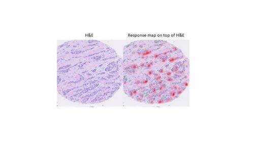
The new technology, which is expected to significantly improve the personalization of cancer treatments, is based on deep learning. It extracts molecular information from biopsy images that have undergone hematoxylin and eosin (H&E) staining - a common staining used to examine tissue taken in a biopsy. The staining allows the pathologist to identify in the tissue, under the microscope, the type of cancer and its degree of severity. However, the staining alone does not make it possible to identify critical characteristics that are essential in determining the appropriate treatment, for example the molecular profile of the tumor, the biological pathways that occur in it, the genetic code of the cancer cells and the common receptors on the cell membrane. The mapping of the receptors on the cell membrane is particularly relevant for personalized medicine; It makes it possible to adapt to cancer patients a treatment that blocks the receptors and inhibits the development of the cancer tumor.
The conceptual innovation of the Technion researchers is in extracting the molecular information from the shape of the cells and their environment (the morphology of the tissue) as it is reflected in H&E scans. According to Shamai and Prof. Kimmel, "pathologists we spoke with said it was an impossible task. This is because a human pathologist cannot infer the properties of a tumor from its shape because of the huge amount of variables. The good news is that artificial intelligence technologies and especially deep learning are able to do this. The computer, as opposed to the pathologist and even the tortuousAnd the most skilled, can characterize the cancer with the help of a complex analysis of its morphology."
Indeed, with the help of image processing and artificial intelligence tools, the researchers showed, for the first time, the possibility of predicting the molecular profile of the cells from the tumor's morphology, that is, just from looking at the tissue as it appears in the standard scans (H&E). "We were able to identify the 'signature' that the cancer leaves in the tissue," explains Shamai. "This is a morphological (formal) signature from which we manage to extract a lot of critical information through our technology. It is important to note that deep learning systems require a huge amount of information, and obtaining the required type of information is not easy. For this purpose, we wrote software code to scan online sources and automatically download thousands of biopsy samples and the relevant medical information approved for research use."
In the study, more than 20,000 scans from 5,356 breast cancer patients were examined. Using the new technology, the researchers were able to map, among other things, estrogen and progesterone receptors from the scans alone and based on the morphology of the cells. As mentioned, the study focused on breast cancer, but the researchers clarify that this is proof of feasibility relevant to all types of cancer. According to Prof. Kimmel, "We were able to show that cancer has a unique signature in the morphology of the tissue and that computerized mapping of this morphology can give us enormous relevant information about the characteristics of the tumor. In the first phase, we estimate that it will be an aid that will help doctors make decisions, and will later be developed into a real clinical tool."
The research was supported by the Ministry of Science and Technology, the National Science Foundation, the Lori Lockey Interdisciplinary Center for Life Sciences and Engineering and Schmidt Futures.
More of the topic in Hayadan:
- Artificial intelligence will make it possible to improve the quality of medical care, already in the near future
- Chapter 12: Artificial life from the book of Kisham Azgad "Life Begins Here"
- Artificial intelligence has contributed to the increase in finding a match for experimental breast cancer treatments

One response
Wonderful