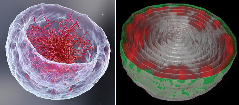
Contrary to the image they got, the DNA molecules in the cell nucleus do not float in the center and do not fill most of its volume

If you had the opportunity to open a biology textbook and flip through the chapters describing how DNA is organized in the cell nucleus, you may have been attacked by a slight feeling of hunger: the image of the DNA chains in the nucleus - long threads floating in liquid - may remind some of us of a rich bowl of ramen or chicken soup With a generous amount of noodles. Soon we will probably have to abandon the "ramen bowl" model - As tempting as it may be - this is what two new studies by Weizmann Institute of Science scientists show. This is not just a graphic presentation, the spatial arrangement of the DNA in the nucleus is essential for understanding genetic expression and control processes - and what may happen when these do not function properly. The studies are the result of close cooperation between the experimental group of Prof. Talila Volk from the Department of Molecular Genetics and the theoretical research group of Prof. Samuel (Sam) Shaffern from the Department of Chemical and Biological Physics.
It all started when Prof. Wolk studied how mechanical forces affect cell nuclei in muscle tissue and found evidence that the contractions (and relaxation) of the muscles have an immediate effect on the expression patterns of genes. "Unfortunately, we could not delve into this intriguing finding as we wanted, since the methods for studying the cell nucleus rely on chemical fixation of the cells - something that impairs the ability to find out what actually happens in active and contracting muscle cells," says Prof. Volk.
To answer this issue, Dr. Dana Lorber from Prof. Wolk's laboratory designed a device that would allow access to the nuclei of muscle cells in live larvae of fruit flies: the tiny, transparent maggot is held in a kind of millimeter slot that allows it to contract and relax its muscles while a powerful fluorescent microscope scans the Taya In this way, the researchers were able to photograph the cell nuclei and their contents - long DNA molecules and supporting proteins organized together in a structure called chromatin.
While expecting to see DNA organized in the cell nucleus like noodles in a bowl of ramen, Dr. Lorber and Dr. Daria Amiad-Pavlov, a postdoctoral researcher in Prof. Wolk's group, got a surprise: instead of the long chromatin molecules, the "noodles", will fill most of the volume of the nucleus, they arranged themselves in a relatively thin layer adjacent to the inner wall of the membrane of the cell nucleus. Similar to the meeting between water and oil, which produces a phenomenon known as "phase separation", the chromatin "separated" itself from the liquid of the cell nucleus and settled in the walls, while the center of the nucleus remained liquid. "Our findings were so surprising and unexpected that for a considerable period of time we tested ourselves again and again and repeated the experiment many times," explains Dr. Lorber.
To better explain the findings, the female researchers from Prof. Shafran's group, who together with the post-doctoral researcher, Dr. Gaurav Bajpai, developed A theoretical model which weighs all the physical forces involved in arranging the chromatin in the nucleus. This model was able to predict what Dr. Lorber and Dr. Amiad-Pavlov saw through the eyepiece of the microscope - the chromatin in the nucleus may separate from the cell's nuclear fluid and settle in the walls depending on the amount of fluid in the nucleus.
So where did the ramen bowl model come from in the textbooks? "When scientists prepare a sample of cells to study their nuclei under the microscope, they actually flatten them and thus change their volume. This change may interfere with the mechanical forces that dictate the way the chromatin is organized, as well as reduce the distance between the top of the nucleus and the bottom," explains Prof. Shafran.
To make sure that the findings are not characteristic of muscle cells of fruit fly larvae only, Dr. Lorber and Dr. Amiad-Pavlov teamed up with Dr. Francesco Roncto, from the laboratory of Prof. Ronan Alon from the Department of Immunology, and together they examined the phenomenon in live white blood cells as well originating from humans. Similar to the cell nuclei in the fruit fly, in these cell nuclei the chromatin was organized in walls. "From this finding it appears that we have apparently uncovered a general biological phenomenon and that this arrangement of the chromatin is evolutionarily conserved - from flies to humans," adds Dr. Amiad-Pavlov.
These findings open new and diverse avenues of research: does the organization of the DNA in the nucleus change in disease states - and is it possible to develop new methods for the early diagnosis of diseases based on this knowledge? Do and how do the mechanical forces acting on the arrangement of DNA in the nucleus also affect cell differentiation during embryonic development? In addition, it is known that the degree of stiffness of the surfaces on which cells intended for research are placed may affect their gene expression patterns. The new findings show that the reason for this lies in the change in the volume of the cell and the nucleus and the forces exerted on them. A better understanding of the array of these forces and their effect on genetic expression patterns may help in tissue engineering.
Dr. Adriana Reuveni from Prof. Talila Volk's laboratory in the Department of Molecular Genetics also participated in the experimental study.
