By creating artificial embryonic and placental stem cells and comparing them, the researchers from the Hebrew University identified about 14,000 sites in the genome that control the development of all the organs of the fetus
A study conducted by a team of researchers from the Hebrew University and published in the prestigious journal Nature Communications led by Prof. Yossi Boganim from the Institute of Medical Research in the University's Faculty of Medicine and Prof. Tomi Kaplan from the School of Engineering and Computer Science and the Department of Computational Biology discovered approximately 14,000 regions in DNA that constitute the initial program most to create an embryo.
Already in 2006, Japanese scientists found that it is possible to take skin cells, insert four embryonic genes into them and reprogram them to behave and function like stem cells that form the embryo itself, which are formed a few days after fertilization. These cells created from the skin cells are artificial embryonic stem cells identical to natural embryonic stem cells, which are in the earliest state in the process of embryonic development and are responsible for the development of all fetal cells, but without the ability to create extracorporeal tissues such as the placenta.
In 2015 Prof. Buganim and his team for the first time cracked the way to produce artificial placental stem cells from skin cells and thus developed the ability to artificially produce the two most primary types of stem cells in embryonic development that are created after the fertilization of an egg by a sperm cell.
The research team from the Hebrew University, which also included to prof' Bogans וProf. Kaplan the PhD students Muhammad Jaber, Ahmed Radwan and Nathaniel Loefer Compare and check what are the processes required for a skin cell to convert its identity and become an artificial fetal or placental stem cell. Prof. Boganim explained: "We used the most advanced analysis methods and examined throughout the transformation process all the changes that the skin cell goes through in order to change its identity to the two types of cells. These analyzes included the changes in the gene expression of the skin cell throughout the process, the changes in the structure and packaging of the DNA within the nucleus of the changing skin cell, as well as changes in the epigenetic marking (chemical molecules that decorate the DNA and are responsible for the proper expression of genes), which were all critical to the success The conversion of skin cells into artificial fetal or placental stem cells."
The researchers discovered that the changes that the skin cell goes through to become an artificial embryonic stem cell or an artificial placental stem cell differ from each other in a sweeping way and at all levels tested even though both processes started from the same skin cells.
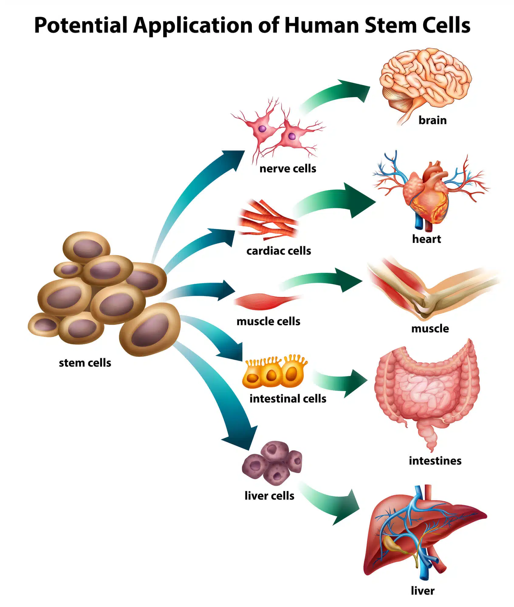
During the transformation of the skin cell into an artificial embryonic stem cell, regions of the DNA important for the formation of the brain, heart and liver began to reorganize within the changed skin cell and prepared it for the ability to differentiate, given an appropriate signal, into brain, heart and liver cells. Alternatively, regions in the DNA of a skin cell that started the transformation process into an artificial placental stem cell began to reorganize in order to give the transformed cell the ability to root and attract blood vessels, as happens during the natural implantation of the embryo.
"The most amazing discovery was observed when we simultaneously compared the two processes and looked at a special chemical molecule called methyl that decorates specific regions of the DNA and is responsible for silencing them," Boganim continues. "We discovered about 14,000 methyl-decorated DNA sites that are found only in the artificial placental stem cells and not at all in the artificial embryonic stem cells. In the next step, we tried to understand the meaning of those sites and were amazed to discover that these sites are responsible for the creation of all the organs and cells of the developing embryo, from the brain, heart, liver and kidneys to the skeleton, spine and connective tissues."
Boganim adds that "this discovery is of enormous importance because it may explain the defense mechanism of the fetus that does not allow early placental cells to develop into fetal cells. Since placental cells are exposed in many ways to damage and infections, there is a protective mechanism that will not allow a damaged cell to migrate towards the developing fetus and integrate into it."
In recent years, a large number of attempts have been made to understand how the stem cells of the fetus and the placenta develop simultaneously but do not fuse with each other. The discovery by Prof. Buganim and his team, which could only be discovered when creating artificial embryonic and placental stem cells, and not by looking at a natural embryo, may shed light on this defense mechanism. The research was supported by the European Union Competitive Research Grants (ERC), and the American HHMI and National Science Foundation grant.
More of the topic in Hayadan:
