How do the cells of the immune system know how to brake quickly?
The cells of the immune system move through the blood vessels at breakneck speed, but know how to brake when needed with a level of precision that would not shame an aircraft landing on an aircraft carrier. This miraculous ability allows them to stop at a precise point on the blood vessel wall to penetrate damaged tissue or the lymph nodes in the body in search of signs of infection. How do the cells manage to identify the breakpoints with such extraordinary precision? Weizmann Institute of Science scientists set out to investigate the issue andFound Because essential immune cells, called T cells, are equipped with braking equipment that is literally at their fingertips.
We tend to imagine cells as smooth spheres, but the surface of many of them - and certainly of all immune system cells - is not smooth at all. On their outer membrane there are flexible protrusions similar to tiny fingers, which help them sense the environment and communicate with it. In order to cross the blood vessel wall and penetrate the lymph node, the "fingers" of the T type immune system cells bind to special proteins found on the wall. After attachment, the cells roll along the wall until their receptors, called CCR7, encounter the chemokines of the lymph node. These small molecules inform the T cells: "Welcome, you have reached your destination". From there, the message passes further inside the cell and activates the LFA-1 protein that binds to specific sticky molecules on the blood vessel wall. Thus the cell stops with a screech of brakes, exits the blood vessel and enters the lymph node, where it performs a scan that lasts from a few minutes to several hours, and if it does not find a foreign factor - it returns to the blood circulation as it came; Thousands of such scans are performed every minute in the hundreds of lymph nodes in the body to ensure our health.

In a previous study, Prof. Ronan Alon and the members of his group in the Department of Immunology that the inhibition signal is generated by the fingers of T cells in less than half a second. This speed is particularly surprising, since a T-cell rolls every second a distance of about 0.1 millimeter, that is, approximately 15 times its diameter - a rate that corresponds in the macro world to the speed of a fighter plane during the landing stages. But when a plane lands on a narrow runway, steel cables designed to stop it are already deployed and ready for action. In contrast, when a T-cell "lands", it seems to have to assemble the landing gear at its fingertips every time. Furthermore, it was previously discovered that the LFA-1 protein - essential for inhibition - is mostly hidden on the cell body, away from the tips of the tiny fingers that cover its surface.

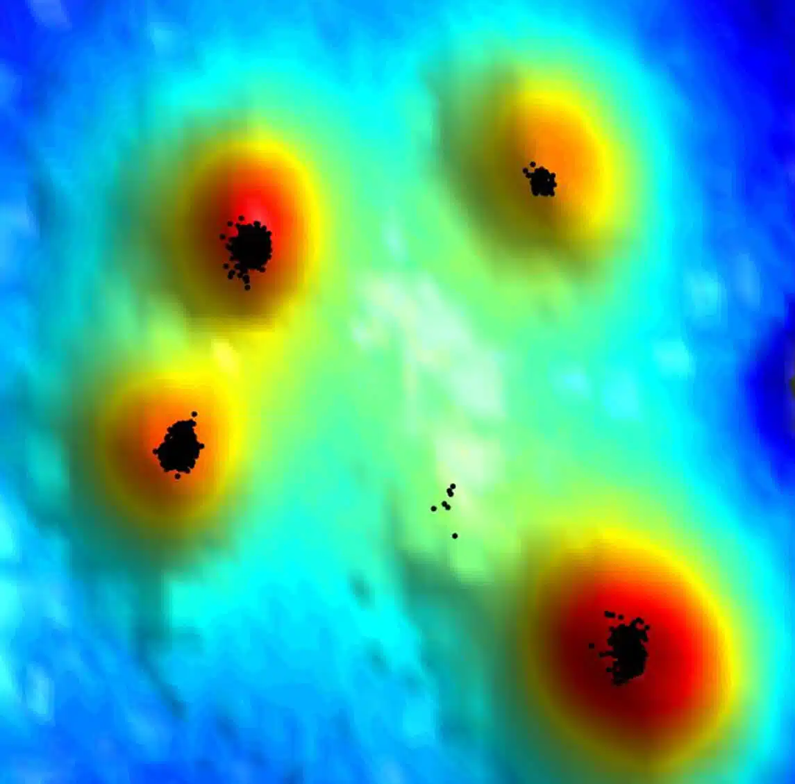
To understand how the T cell manages to assemble its braking equipment at record speed, Prof. Alon teamed up with Prof. Gilad Haran from the Department of Chemical and Biological Physics, who developed a new approach to studying the surface of cells at the level of single molecules using super-resolution microscopy. It is a combination of two technologies: one method for determining the location of different molecules using fluorescent markers, and another method for creating a sort of topographical map of the surface of the cell, including its fingers, based on the intensity of the fluorescent signal. "We map the hills and valleys on the surface of the cell and create a topographic map similar to the one that shows the shape of the Earth's surface," explains Prof. Haran.
"All the equipment needed for rapid braking is ready to be assembled at the fingertips of T cells. This way the cells can produce the braking signal in less than half a second"
Dr. Shirsando Ghosh, a postdoctoral researcher in Prof. Haran's lab, and Dr. Sarah Feigelson from Prof. Alon's lab, led the new study, in which the scientists discovered how T cells perform their stunt. They found that CCR7 receptors are concentrated in the most accessible part - exactly at the fingertips of the cells. It was also discovered that the LFA-1 molecules are mostly found on the cell body, but about 5% of them are on the surface of the fingers near CCR7. Another and no less important thing: all the auxiliary molecules needed to transmit the braking signals were also found at the fingertips, in close proximity to the CCR7 receptors. "All the equipment needed for rapid braking is ready to be assembled at the fingertips of T cells. This way the cells can produce the braking signal in less than half a second," says Prof. Alon.
Apart from revealing the modes of action of the immune system, the research findings also open the door to the study of the mechanisms that control the movement of different cells, including cancer cells that may drift in the blood circulation and stop in different places for the purpose of creating metastases. The findings may also help to develop ways to control the movement of the cells of the immune system: in this way, it will be possible, for example, to increase the number of CCR7 receptors in the fingers of the cells through genetic engineering in order to accelerate their arrival in the lymph nodes and thus increase the effectiveness of various vaccines, or, alternatively, to restrain the movement of Harmful immune cells at work in autoimmune diseases.
Dr. Alessio Montresor and Prof. Carlo Laudana from the University of Verona in Italy participated in the study; Dr. Eyal Shimoni from the Department of Chemical Research Infrastructures at the Weizmann Institute of Science; Dr. Francesco Roncto from the Department of Immunology of the Institute and Prof. Daniel Legler from the Institute of Biotechnology Thurgau, Switzerland, belonging to the University of Konstanz.
More of the topic in Hayadan:
