With the help of an innovative development by university researchers, doctors will for the first time be able to measure the size of the pupil and its reaction to light even when the eyes are closed: "The development will enable early identification of abnormal parameters and will contribute to saving the lives of patients with severe brain injuries"

Pupil measurement is a key component in the neurological assessment of patients hospitalized in a coma, for example after a stroke or head injury. Asymmetry in the size of the pupils, or their lack of response to light, may indicate a life-threatening increase in intracranial pressure, which requires immediate medical intervention. Today, in intensive care units, measuring the pupils of patients who are in a coma is done manually and every hour or so - a brother or sister opens the patient's eyelid, measures the size of the pupil with a ruler, and shines a flashlight to check if the pupil reacts to light. The new technology developed, will allow for the first time to collect this information automatically and make it significantly easier for the medical teams.
The technology was developed by a team led by Dr. Tzevi Batos and Dr. Amnon Buxboim, from the School of Engineering and Computer Science, the Institute of Life Sciences and Bioengineering Center of the Hebrew University and Prof. Jose Cohen, a neurosurgeon from the Hadassah Ein-Karem Hospital and will enable continuous monitoring of the pupils without contact with the patient, and immediate notification to the treating team in the event of life-threatening abnormal findings. The idea for the project was born in the entrepreneurship course Bio-Design of the Department of Bioengineering at the Hebrew University. In the course, integrated teams of doctors and students from several fields are formed: engineering, law and economics, whose goal is to characterize the need for new biomedical devices, conduct market research and propose devices that will meet the medical need.
The device developed by the researchers shines infrared light through the temple area. Dr. Batos explains that "the technology is based on the principle that light at certain wavelengths, for example in the infrared range, can pass through tissue. The device we developed illuminates with light that penetrates the eyeball, disperses inside it, and part of it exits through the pupil and out through the eyelid." The team of researchers placed an infrared-sensitive camera in front of the eye, and managed to photograph the spot of light passing through the pupil.
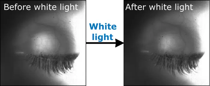
To measure the contraction of the pupil, the researchers used the light transmission properties of the tissue, but in the opposite direction: in the same way that we are able to sense sunlight even with closed eyelids, the researchers shone white light on the eye from the front, which passed through the eyelid and the pupil, and was seen by the retina as red light. The light caused the pupil to contract, which the researchers were able to measure with the help of another infrared photograph. Since then, a first clinical trial was carried out in healthy volunteers, in which they were able to perform the measurement in 38 out of 39 volunteers, proving the feasibility and safety of the new method.
As a result of the research, a company was established that continues the development of a device based on the same technology that will allow the measurement of two eyes at the same time in unconscious patients in a supine position. Prof. Cohen concludes that "the development of such a device for continuous monitoring will enable the early identification of abnormal pupil parameters and thus contribute to saving the lives of adults and children with brain injuries".
More of the topic in Hayadan:
