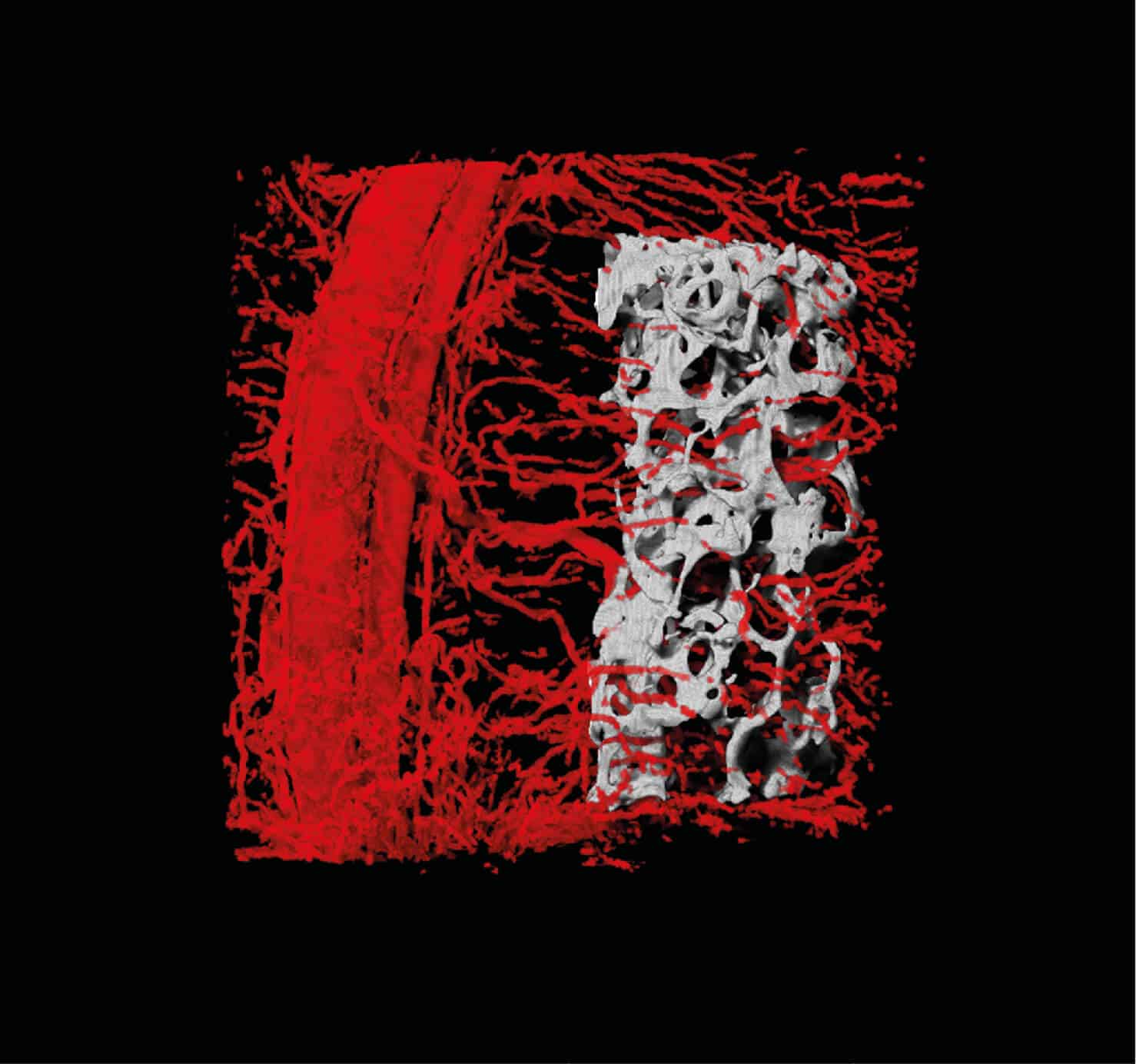
A technology developed at the Technion makes it possible to grow bone tissue networked with blood vessels in the laboratory. This achievement accelerates the absorption of the engineered tissue in the affected organ

Bone restoration using engineered tissue: researchers at the Faculty of Biomedical Engineering at the Technion present in the journal Advanced Functional Materials Success in creating bone tissue containing a network of blood vessels. In an experiment with rats, the tissue graft was successfully absorbed into the damaged bone. The research was led by Prof. Shulamit Levenberg and Dr. Idan Radansky.
Various injuries in our body heal naturally - scratches heal, wounds close and even internal damage sometimes recovers without external help; However, in many cases, for example in the case of trauma or the removal of cancerous tissue, the damage is too severe for the body to overcome without medical intervention.
One of the ways to restore damaged tissue is an autograft - transplanting tissue taken from a less vital organ in the patient's body. For example, in the case of significant tissue loss in the mouth and jaw, it is now possible to harvest a combined tissue (bone and soft tissue) from the leg and reimplant it in the damaged area. Transplantation from one's own source has a certain advantage - the body does not reject the transplant as it does in the case of a donation from a foreign person - but there are also many disadvantages to this procedure, chief among which is damage to the organ from which the tissue is harvested. That is why there has been a scientific race for alternative solutions for many years, and one of the main ones is the creation of a laboratory-engineered suspension (implant).
Easy to talk - hard to do. For the rack to be effective it is not enough that it be precise in terms of its shape and mechanical properties; It should integrate into the target organ from a biological point of view as well and become part of it - one living tissue. This is where Prof. Levenberg's many years of experience in tissue engineering and Dr. Redansky's familiarity with oral and maxillofacial surgery come into play - knowledge he acquired during his doctoral studies in dentistry.
Prof. Levenberg entered the world of tissue engineering during her postdoctoral period at MIT, where she developed with Prof. Robert Langer the first engineered muscle that contained blood vessels. Since then, she has been researching and developing new technologies to create three-dimensional tissues for transplantation that will be absorbed quickly and effectively in the target organ. In recent years, she has recorded a series of scientific achievements, including improving the formation of blood vessels in the engineered tissue - an improvement that accelerates the absorption of the tissue in the body - and the rehabilitation of a damaged spinal cord using engineered tissue. In addition, she opened the bioprinting center at the Technion, harnessing XNUMXD printing to create tissues for transplantation.
Dr. Idan Redansky has been leading a relatively new direction in the Levenberg laboratory for the past five years - breeding (in the laboratory) of tissue Combined containing muscle and bone And no less important - you the vascular system which is essential for the future absorption of the engineered bone in the body.
Creating blood vessels in engineered tissue is already a difficult challenge, but it is twice as difficult in bone tissue due to its density and stiffness. The solution developed by the Technion researchers is based on coupling between the bone and soft tissue that sends the blood vessels into the bone. To examine the applicability of the technology in the clinical aspect, the bone was implanted with the blood vessels in the body of the model animal and indeed, the blood vessels of the transplanted bone united with the blood vessels of the damaged organ, the scaffold was embedded in the target tissue andWithin a few weeks a full recovery was achieved.
"Idan's research expresses the unique combination that the faculty promotes - a combination of engineering and medicine," says Prof. Levenberg. "He harnessed a deep understanding of biological processes, and all the knowledge he acquired in the studies of surgery and dentistry, to the development of a new technique for restoring damaged tissues. There was also vital and useful cooperation between various faculties at the Technion, between Israel and abroad and between the Academy of Industry: we received the bone grafts from Prof. Gordana Vaniak-Novkovich of Columbia University, and the experts of the Bruker-Skyscan company helped us a lot with the imaging of the engineered blood vessels and the bone tissue inside the regenerating tissues. This collaboration, with the generous assistance of the European Research Fund (ERC) and the National Science Foundation (ISF), allowed us to register this achievement, which we intend to continue to develop for the benefit of human transplantation."
At the end of his doctorate, Dr. Radansky plans to return to the clinic and start the oral and maxillofacial surgery residency program with a research orientation at the Galilee Medical Center, under the direction of Prof. Samer Srouji. There he hopes to apply the achievements of current research and implement solutions from the field of tissue engineering to contribute to patient care. According to him, "Injuries to the jaw dramatically affect the patients' quality of life. Printing titanium implants is a useful interim solution with reasonable results, but with all the technological advances, titanium allows Mechanical fusion only between the implant and the damaged organ, without biological assimilation. This is where we come into the picture, and our ambition is to develop in the laboratory a complete bone that will replace the jaw in all its components. A lot of work is required here, but our research shows that we are on the right track, and it is relevant not only for jaw reconstruction but for any surgical process that involves bone and soft tissue."
The research was supported by ERC (European Research Commission, Horizon 2020 program), ISF (National Science Foundation) and NIH (US Institutes of Health) grants.
for the article in Advanced Functional Materials click here
More of the topic in Hayadan:
- The google maps of the brain: locating RNA fragments in brain cells without removing the tissue
- Rapid development of medicines with the help of bionic chips, without the need for animal experiments
- Each and every cell is stressed: first mapping of the "stress axis" at the individual cell level
- Prof. Shulamit Levenberg of the Technion developed a method that accelerates the absorption of transplanted tissues in the body
- A new tissue printing center was inaugurated at the Technion

2 תגובות
If it is possible to grow meat for food then it is also possible to grow bone and muscle using a personal and adapted source.
Great progress. But is it really impossible to do without what you call in despicable insensitivity the "model animal"?
Did they make sure it wouldn't hurt her? That you won't be terrified of the whole process?
...from my experience in laboratories, nobody cares - only the article is important, and the animal (a creature with awareness and emotions and fear and pain) is thrown into the garbage, without even being put to sleep first.