Researchers from the Weizmann Institute have deciphered how scallops see in the water using 200 tiny blue eyes

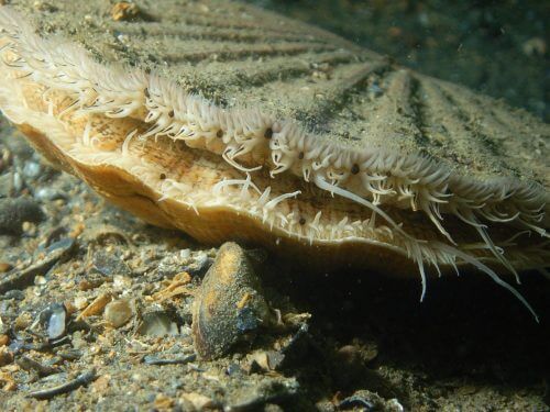
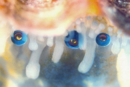
Scallops see with the help of 200 tiny eyes that surround their body. Instead of focusing light through a lens, the oysters' eyes consist of a mirror at the back which reflects light onto two different retinas. Scientists from the Weizmann Institute and their research partners at Lund University in Sweden described recently in detail the structure of this mirror and its optical properties in the scientific journal Science.
Scallop oysters live on the bottom of the ocean, and unlike other oysters, they can swim. Their glowing blue eyes are located on the upper and lower edges of their round bodies. Dr. Benjamin Palmer, a post-doctoral researcher in the research groups of Prof. Leah Eddy and Prof. Steve Weiner From the Department of Structural Biology, it is said that to explain how the oysters' unique vision system works - from the nanometer to the millimeter point of view - collaboration between physicists, chemists and biologists is required who have combined advanced research methods with simulations of various types.
From the nanometer perspective, the mirror at the back of the eye is made of tiny crystals of guanine. Prof. Addi, Prof. Weiner and others have previously shown that when these transparent crystals are organized in a multi-layered structure, they are able to reflect light; They discovered this property in silvery fish scales, the protective covering of spiders, and the outer layers of crustaceans. Using an electron microscope, the researchers showed that in the eye mirrors of the scallops, the crystals are nearly perfect squares that tile the surface of the mirror. The research team analyzed the tiles in detail, revealing their optical properties, as well as the way the material's nanoscale structure creates an effective light-reflecting surface. "The tiles are arranged similarly to the small mirrors that make up the mirror of a large reflecting telescope," says Dr. Palmer. Prof. adds Dan Oron, from the Department of Physics of Complex Systems: "The square tiles in the eyes of oysters are organized in such a density that leaves no gaps that could create a lack of focus of light rays in the image they reflect."
"Each of the eyes sees real shapes - and not just light and shadow like in many other marine creatures," says Prof. Addi. "They can clearly distinguish the shape of a starfish that may approach to devour them"

The team discovered that the multi-layered structure of the mirror effectively reflects the wavelengths of light penetrating the oysters' habitat - with peak reflections in the blue range of wavelengths.
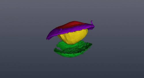
The group then turned to investigate the three-dimensional structure of oyster eyes. Together with Dr. Gavin Taylor from Lund University, they examined what happens to a light beam that passes through the thin and dysfunctional lens, the inner eye and both retinas, and is then reflected back from the mirror to the retinas. The simulations that followed the course of the light beam showed that light from different entrance angles is reflected in a focused manner from different parts of the convex surface of the mirror to one of the two retinas. This analysis suggests that one of the two retinas may be used for peripheral vision in dim light, while the other is better focused in the central line of sight of the eye. Further simulations and experiments revealed that this vision system is an effective solution for producing a relatively focused image in the oyster's compact eye.
"Each of the eyes sees real shapes - and not just light and shadow like in many other marine creatures," says Prof. Addi. "They can clearly distinguish the shape of a starfish that may approach to devour them."

"We saw a very high level of control in directing the growth of the guanine crystals," adds Prof. Weiner. "The small dimensions of the mirrors leave very little room for errors in their curvature."
"We still don't know exactly what the scallop sees through its 200 eyes. The so-called oyster does not have a brain that can process the images - but there is evidence of some coordination between its many eyes"
For the researchers, it is not surprising that such an unusual vision system developed in the ocean. Sight first appeared in ocean creatures, and evolved into several unique forms. "It is much more difficult to see under water than in the air," explains Prof. Oron, "lenses may be, in the end, more efficient for focusing light, but the mirrors in the eyes of oysters work surprisingly well."

"We still don't know exactly what the scallop sees through its 200 eyes," says Prof. Addi, "the so-called scallop does not have a brain that can process the images. But there is evidence of some coordination between her many eyes."
"Besides revealing the workings of this fascinating biological optical system," says Dr. Palmer, "the research was unique in the way it wove the 'story' from different perspectives and methods. It was impossible to understand how the system works without the contribution of each of us."
Dr. Vlad Brumfeld and Dr. Nadav Elad from the Department of Chemical Research Infrastructures, Dr. Dvir Gur from the Department of Molecular Biology of the Cell and Physics of Complex Systems, Dr. Michal Shemesh from the Department of Structural Biology and the Department of Molecular Biology of the Cell, and research student Aya Usherov from the Department of Materials and Surfaces.
