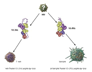
How do two almost identical viral envelope proteins bind to different receptors found on different cells in the human body?

The two main instances of 1-HIV (the strain responsible for 90% of AIDS cases) are called R5 and X4. They differ from each other to a significant extent, both in the type of immune system cells they attack, and in the stages of the disease they cause. The R5 virus binds to molecules (secondary receptors) on the membranes of immune system cells called macrophages. In doing so, it causes the weakening of the entire immune system, in the "dormant" phase of the syndrome (the carrier phase). In contrast, X4 binds to secondary receptors found on the membranes of T-cells, causing the rapid death of these cells. Following this, the fatal phase of the syndrome erupts, in which a complete collapse of the immune system occurs. Despite the great functional difference between these two viral versions, the difference between them can be due to a change in only one or two amino acids in a certain region of the virus's envelope protein. Because of this, the frequent genetic changes that occur in the virus can cause it to switch from the R5 form to the X4 form. In other words, the mutations that occur in the virus, while inside the human body, greatly influence the development of the disease, and allow the virus to diversify its attack strategies.
How do two almost identical viral envelope proteins bind to different receptors found on different cells in the human body? Why does a change in a single amino acid so greatly affect the virus's attack mechanisms? Prof. Jacob Englister and research students Esnat Rosen and Michal Sharon, from the Department of Structural Biology at the Weizmann Institute of Science, used nuclear magnetic resonance (NMR) to closely examine the difference between the virus variants. This difference is due to changes in the three-dimensional spatial structure of a certain segment in the envelope proteins. This section, called V3, determines the type of appearance of the virus, and is involved in its binding to the secondary receptor on the target cells - an essential step in infecting the cells with the virus (binding to the secondary receptor CXCR-4 causes infection of T-cells, while binding to the 5-CCR receptor causes infection of macrophages ).
The scientists synthetically created the sequence of amino acids that make up the V3 segment of the two versions of the viral envelope protein, and examined the structure formed when the different versions bind to two different antibodies (the first antibody was obtained from a mouse vaccine with the envelope protein of the X4 virus and neutralizes only a special strain of X4, and the second was obtained from an HIV-1 carrier and neutralizes a wide variety of R5 and X4 strains). The researchers assumed that when a segment of the viral envelope protein binds to an antibody, it assumes its natural spatial form, the one that exists in the free virus and allows it to bind to the various receptors of human cells. At first glance it seemed that, when bound to the antibody, the two versions of the V3 segment of the viral envelope protein form a horseshoe-like spatial pattern. But, a careful examination revealed differences between the two versions: the arrangement of the amino acids on one side of the horseshoe is the same in both versions, while on the other side there is a shift of the amino acids in the envelope protein of a virus from the X4 version compared to the position of the corresponding amino acids in the virus from the R5 version. As a result of the shift in the position of the amino acids, the position of the hydrogen bonds between the amino acids on either side of the horseshoe also changes. Another change: the side chains of the amino acids on one of the sides of the horseshoe in the envelope protein of the X4 version of the virus face in the opposite direction compared to their orientation in the R5 version of the virus. This creates, in fact, significant differences in the shape of the viral envelope protein and in the spatial organization of the residues that form its binding site. These differences are the reason why viruses of different variants bind to different secondary receptors displayed on the membranes of different cells, thus attacking the immune system in different ways. But what is the exact relationship between the changes in the chemical sequence of the viral envelope protein and the changes in its structure? And why does replacing a single amino acid affect the protein structure so dramatically?

