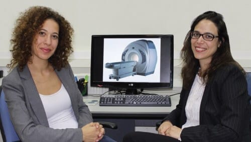
Ahead of the MIXiii 2014 conference and exhibition, the major annual event of the Israeli high-tech and biomed industries, which will be held this May, we interviewed Dr. Efrat Sasson and Dr. Tamar Blumenfeld-Katzir, from BioImage. In the interview, they tell how imaging technologies are currently being mobilized for the biomedical research service, what is the connection between bypass surgery in the heart and the brain, and how all this connects to advanced rehabilitation technologies
Basic biomedical studies mainly include clinical assessment of the disease and histological or biochemical measurements of the tissue. However, today more specific methods are at the forefront of technology. Various imaging methods, among them MRI, enable high-resolution three-dimensional tissue imaging, and provide diverse quantitative, structural and functional parameters.
What are those advanced imaging methods? And how do these imaging methods fit into medical research? It is widely believed that medical imaging images are 'a simple photograph of body tissue'. “New imaging technologies are like a kaleidoscope; With them you can look at the same organ in several ways and each time see a different picture. Each type of image highlights different information about the disease," say Dr. Efrat Sasson, and Dr. Tamar Blumenfeld-Katzir, founders of a company bioimage, BioImage)) a company that has been operating for almost five years, which was established with the aim of bringing advanced imaging technologies to the biomedical community.
The company will present its innovations as part of MIXiii 2014, a joint event for Israel's hi-tech and biomed industries, to which the chief scientist from the Ministry of Economy is also a member - which will be held at the Tel Aviv Exhibition Center between May 20-22, 2014. The event will give visitors the opportunity to witness up close the potential inherent in Israel's advanced technological industries, Participate in discussions and panels, watch live demonstrations and find business partnerships.
Using the advanced imaging methods, it is possible to obtain a variety of biomarkers for characterizing tissues and diseases and for evaluating the effectiveness of drugs or different treatments, say Dr. Sasson and Dr. Blumenfeld-Katzir, who completed a doctoral course at the University of Neuroscience and MRI and in the project which they established now bring knowledge from the academy to promote biomedical research in the way of bringing innovative treatments to the world of medicine in hospitals. Among its clients, leading companies in the economy in various fields and academic researchers in the fields of neurology, psychiatry, orthopedics, heart, cancer and more.
Risk to brain tissue
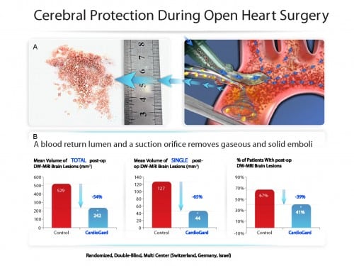
One of the examples presented as part of the MIXiii 2014, is a study carried out by the Bioimage company, to characterize brain lesions after open heart surgery, for the Cardioguard company and in collaboration with Prof. Gil Bolotin and Dr. Ayelet Eran from Rambam Hospital.
Bypass surgery, which is open heart surgery, is considered a common surgery and almost risk-free. However, open heart surgery poses a risk to brain tissue. A large proportion of those who undergo open heart surgery experience a cognitive decline, some of which are transient and in rare cases a stroke can even occur. What is the connection between bypass surgery in the heart and the brain? The reason for this is the appearance of a large number of emboli particles that are released from the blood vessels during the operation, the emboli particles are high in fat and may reach cerebral blood vessels and clog them. The Israeli Cardioguard Medical has developed a unique tube - a cannula - which simultaneously allows the suction of the emboli and the free flow of the blood during the operation (photo 2A). In order to prove the effectiveness and safety of the product, a study was carried out at several sites in the world under the leadership of Prof. Bolotin, director of the heart surgery department at Rambam Hospital and in collaboration with Bioimage.
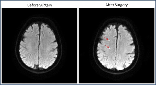
The study was conducted in four medical centers - in Israel, Rambam Hospital, Switzerland and Germany. In half of the heart surgeries in the study, Cardioguard Medical's unique tube was used. The patients underwent a brain MRI scan before and after the operation, using an advanced method based on the mobility of water molecules. Brain areas where there is a decrease in blood supply due to the embolisms, enter "distress" and swell, which is seen in the MRI scan as bright spots (photo 3). Bioimage, in collaboration with Dr. Ayelet Eran from the neuroradiology unit at Rambam, showed that much fewer lesions (bright spots in the brain scan) can be seen in surgeries in which a cannula was used. Lesions were identified in about 40% of the operated on in the group where the cannula was used compared to about 60% of the control group. The Biomag company, in collaboration with Dr. Eran, performed a volumetric measurement of the brain lesions, which allowed us to obtain quantitative information about the damage to the brain tissue. Not only were there fewer lesions following the use of the cannula, the volume of the lesions was significantly lower (Figure 2B).
The knowledge and expertise of Bioimage, in analyzing the results of scans from several centers in the world, was manifested in the verification of the uniformity of the scans between the various centers throughout the project and in a quick and professional response", says Dr. Walid Haddad, CEO of Cardioguard Medical.
Advanced restoration technologies
Another example of a study in which advanced imaging methods provided important information is that of the Israeli company BrainQ Technologies, which develops advanced rehabilitation technologies for various neurological injuries, such as stroke. In a study conducted for them by the Bioimage company, rats were scanned with an MRI machine designed for rats and mice at Tel Aviv University. These are stroke model rats (the model was made by the pharmaseed company) and were treated with BrainQ technology. The rats were scanned at several time points, before, during and after treatment.
"BrainQ has been working with Bioimage for over a year. During this period, Dr. Blumenfed-Katzir and Dr. Sasson performed the simulations in the company's preclinical experiments," says Dr. Yaron Segal, founder of BrainQ. "The company focuses on finding advanced rehabilitation technologies so that the ability to perform internal imaging and collect information from live models throughout the trial period is of utmost importance to us. Tamar and Efrat present a new world to our kind of research and technologies that were open until now only to the academic world. A high-level analysis of the findings and the presentation of the scientific explanation for the phenomena we see, allows us to see the process and prove the assumptions underlying our research and development, and therefore allow us to move more quickly to clinical trials, in which we can prove that the treatment methods that BrainQ develops do indeed help people. "
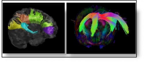
"Since advanced imaging technologies enable impressive visualization and show results relatively quickly compared to traditional methods in research, they can help decision-making processes at important junctures in medical research in the preclinical and clinical stages. For example, restoring fiber systems in the brain can demonstrate a therapeutic effect in various diseases such as multiple sclerosis, brain tumors and stroke, and more _(Photo 4)", say Dr. Sasson and Dr. Blumenfeld-Katzir. "Correct decisions in the early stages of research can save many resources and mark the way for the companies that develop medical devices or medicines. It is possible to obtain convincing and impressive initial results at an early stage and thus help in raising resources for research. In more advanced stages, the use of imaging methods constitutes a significant backup for the clinical results and in some cases is even necessary for proving the effectiveness and safety of the treatment in front of parties such as the FDA".
