Fetal Development

The Balbian body, a unique structure that organizes essential molecules in the early stages of the embryo's development, illustration: Kar at, all

The Libyan Body: Cracking the Secret of Life's Beginning

The researchers used protein staining techniques to reveal the identity of the cells in the organoids they created. In the picture you can see four organoids of the central nervous system in the fetus, where in purple are proteins associated with the development of the front and middle brain, in green - the hindbrain, and in red - the center of the spinal cord

from the brain to the tail

Researchers from the University of Michigan and the Weizmann Institute of Science have for the first time developed a central nervous system on a chip that faithfully simulates that of the human fetus - from the end of the spinal cord to the forebrain
Mouse, rabbit and human embryos (right to left) in the same developmental stage - gastrulation (days 8-7.5 of pregnancy in rabbits and mice and around day 18 of development in humans). At this stage the human, rabbit and other vertebrate embryos are almost flat disc-shaped, while the mouse embryo is elongated cylinder-shaped. The images of the mouse and rabbit embryos were created as part of the current study, the image of the human embryo is taken from a study published in 2021 (Richard CV Tyser et al. Single-cell transcriptomic characterization of a gastrulating human embryo. Nature 600: 285-289)

In the rabbit hole

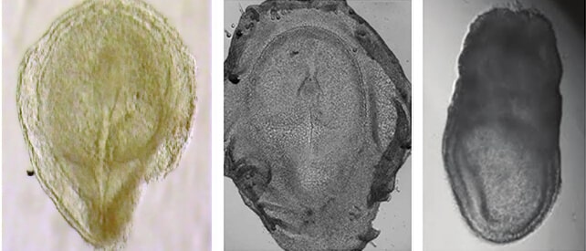
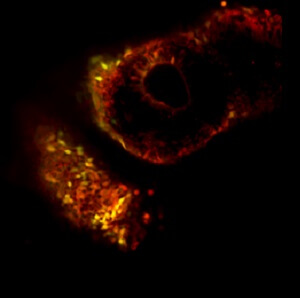
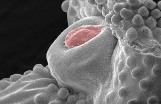
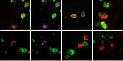
The institute's scientists developed a method that allows real-time monitoring of the development of embryos at the beginning of their journey and applied it for the first time to rabbits. The comparison they made between embryonic development in rabbits and mice gave rise to answers to fascinating questions about
Cells. Illustration: depositphotos.com

Start with one cell

The long and winding road to doubling the amount of biological material available for research
Diagram of the spatial interaction between the DNA control regions and the controlled genes. The "reporters" (in red and green) reveal these interactions. The laboratory of Dr. Yonatan Stanzler, Weizmann Institute

A cell following its fate

Neurons of a mouse embryo grown in a Petri dish. The axons, the extensions of the nerve cells, are the green extensions in the picture. Source: NIH.

A little less, a little more

A centriole as photographed with an electron microscope. Photo: © Pierre Gönczy/EPF

Beyond genes: do centrioles carry biological information?

immune system. Illustration: shutterstock

Get vaccinated very early

Modular model: two different groups of cells - the builders of the cartilage (orange) and the builders of the bone protrusions (green) - are involved in the development of the bone in the fetus

The collagen puzzle

Dr. Yaakov Hana. Photo: Weizmann Institute

A new method for increasing the production efficiency of stem cells

Right: Itamar Harel and Prof. Eldad Tzhor. genetic network

Myocardial

A cock embryo's male genitalia (artificially dyed red) before it withers, as seen under a scanning electron microscope. Figure (AM Herrera and MJ Cohn, University of Florida)

Why did the birds lose their male genitalia?

Embryonic stem cells that develop into gametes, from which the gene encoding the Utx enzyme has been deleted, are labeled with a bright green fluorescent protein. In each of the columns is marked (in red, purple or orange) a gene essential for the survival and development of the gametes. A comparison of the tenth day of cell development (top row) with day 12 (bottom row) shows that the expression of the four essential genes is stopped. As a result, the stem cells do not develop into sperm or egg cells, but die. Photo: Weizmann Institute

back to the Future

From the right: Dr. Rachel Serig, Alina Molchedsky, Dr. Ariel Rinon, Dr. Eldad Tzhor and Prof. Verda Rutter. Rewired

added to his role

The female skeleton - back view. From Wikipedia

bones, muscles and tendons