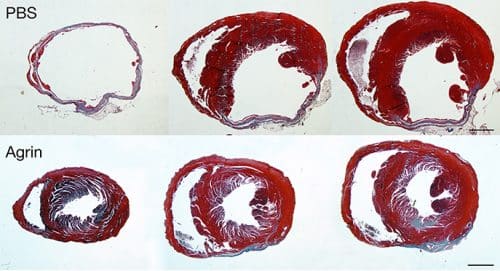
Mice that had a heart attack returned to full cardiac function after an injection of the agrin protein

Heart disease is the leading cause of death in the western world. One bright spot in this matter is the fact that mammals, including humans, have the ability to renew their heart tissue and repair damage themselves - close to birth. The disappointing facts are that shortly after birth, the ability to self-repair disappears, seemingly never again; On the other hand, there are no treatments that help repair damage to the heart muscle. In a new study recently carried out at the Weizmann Institute, scientists discovered, in the hearts of newborn mice, a protein that may restore the ability to regenerate heart tissue. When this protein, called Agrin, is injected into the hearts of adult mice that have suffered a heart attack, it appears that the protein makes it possible to restore the muscle tissue. These findings, which were published in the scientific journal Nature, point to new research directions in the field of cardiac rehabilitation.
Prof. Eldad Tzhor, who led the research with research students Elad Bashet, Alex Ganzlinach, and other researchers in the Department of Molecular Biology of the Cell at the Weizmann Institute of Science, explains that after a heart attack occurs, the body's response to the damage is not effective to the extent necessary to heal the heart. For, as soon as the heart muscle cells, called cardiomyocytes, are damaged, they die and are replaced by scar tissue, which is unable to contract, and therefore cannot participate in the vital pumping action of the heart. This in itself adds to the burden on the muscle tissue that remains normal, and ultimately leads to heart failure.
Regeneration of the heart even in adulthood is a phenomenon that occurs among some vertebrates. Fish, for example, can effectively regenerate a damaged heart. Animals that are evolutionarily closer to us - mice - are born with this ability, but lose it a week after their birth. This week is a window of opportunity for Prof. Tzhor and the members of his research group, who focus on researching the processes that enable the regeneration of the heart.
Prof. Tzhor and Elad Bashet believed that the secret of regeneration lies, in part, outside the heart cells - in the support tissue that surrounds them, known as the extracellular environment or ECM. Many intercellular signals are transmitted through this tissue, while other signals await fitness within its fibrous structure. Therefore, the research team began to examine this environment in newborn and week-old mice, removing the cells from it and leaving only a cell-free tangle of proteins. Next, they added extracellular protein fragments to cardiac tissue cultures. Indeed, they found that the young tangle, as opposed to the more mature one, allowed cardiomyocytes in culture to divide.
After analyzing and scanning the proteins of the extracellular environment, the researchers identified several proteins that may cause this reaction, including the agrin protein. The role of agrin in other tissues was already known in the past, and especially in all that has been said in creating the connection between nerve and muscle, where it helps regulate the signals transmitted from nerve cells to muscles. In the hearts of mice, the level of agrin protein is in decline in the first seven days after birth. Therefore, the scientists tried to find out if the protein affects cell regeneration in the heart, and indeed, the results were impressive: when the researchers added agrin to heart muscle cell cultures, the cells began to divide.
After only one injection of Agrin into the hearts that had seizures, the hearts were almost completely healed, and returned to full function
In the next step, the researchers tested the effect of agrin on models of cardiac injury in mice, and asked to find out if the protein could heal damage caused to the heart tissue. Here, too, the results were not slow to arrive: they discovered that after only one injection of agrin into the hearts that had seizures, the hearts were almost completely healed, and returned to full function. Although the researchers were surprised to discover that a period of time of more than a month was required to notice the full effect on the function of the heart and its regeneration, at the end of the recovery period the scar tissue was significantly reduced, and its place was taken by living tissue that contributed to the heart's pumping action significantly.
Prof. Tzhor hypothesizes that in addition to a certain regeneration of cardiomyocytes, agrin also affects in some way the body's inflammatory and immune response to a heart attack, as well as suppressing the scarring that leads to heart failure. However, the length of the recovery process remains a mystery, since the agrin molecule itself is cleared from the body within a few days after the injection. "Obviously, this molecule creates a chain of events," he says. "We discovered that it binds to a previously unexplored receptor on the surface of heart muscle cells, and this binding returns the cells to a slightly less mature state, closer to that of the fetus, and releases signals that may, among other things, cause cell division." The results of experiments with mice genetically engineered so that the levels of agrin in their hearts were low also support this idea: in the absence of agrin, newborn mice were unable to repair heart tissue after injury.
The research team later showed that agrin has a similar effect on human heart cells grown in culture. Prof. Tzhor and the members of the group are now working to accurately understand the processes that take place in the period of time between the agrin injection and the return to full cardiac function. Also, Prof. Tzhor and Dr. Kafir Baruch Umansky began conducting preclinical studies in pigs in Germany, in collaboration with Prof. Christian Kopat Jermeis from the Technical University of Munich, with the aim of identifying the effect of agrin on repairing cardiac damage.
The findings of this study indicated the important role played by the extracellular environment, both in the maturation of the heart and in promoting its regeneration, and this insight may help in the development of treatment for heart diseases.
Researchers from different parts of the world participated in different phases of the research, in particular Prof. Shenhav Cohen from the Technion and her doctoral student, Yara Eid, who contributed new insights into the biochemical mechanism by which agrin works; Prof. Nenad Bursak from Duke University, North Carolina; James Martin of Baylor College of Medicine, Houston, Texas. Members of the Nancy and Steven Grand National Center for Personalized Medicine and Prof. Irit Sagi from the Department of Biological Control at the Weizmann Institute of Science also participated in the study.
