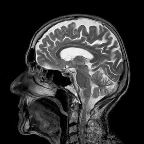
An MRI scanner, based on magnetic resonance technology, has since its invention provided unprecedented diagnostic capabilities in the world of imaging. Recently, a team of researchers from the University of North Carolina in the USA (UNC) took advantage of this technological advance in order to identify signs of autism in the brain of six-month-old children (for the attention of the community opposed to vaccines given at the age of one - A.B.H.).

Autism (Kener syndrome) is a developmental disability resulting from a developmental, hereditary and congenital neurological difference. It is difficult to detect autism at an early stage and sometimes the first signs, such as lack of eye contact, appear only after the age of two. The research team, led by Joseph Piven and Heather Cody Hazlett, conducted the study in four medical centers in the United States, on 109 infants who are at high risk of having autism because their siblings were diagnosed with autism (a 1:5 chance , where the chance in the population is approximately 1:100) and at the same time, on 42 babies without a family background of autism. They examined, with the help of an MRI scanner, the babies' brains while they slept at the ages of 6 months, 12 months and 24 months.
At the age of two years, 15 of the babies who had a high risk of autism due to a family history, were indeed diagnosed with autism (based on failure to make eye contact, delay in the ability to speak and delay in other developmental areas) and the research findings showed that their brain volume increased between a brain MRI scan at age 12 months and an MRI scan brain at the age of 24 months, relatively quickly compared to children without autism. Another finding in the MRI scans that differentiates between the babies diagnosed with autism at the age of two and those who were not diagnosed, is found between a brain MRI scan at the age of 6 months and a brain MRI scan at the age of 12 months, when in the babies diagnosed with autism the surface area of the cerebral cortex (a measure of the size of the cerebral folds in the outer part of the brain), grew faster compared to those who did not receive a diagnosis. In summary, the study's diagnostic algorithm was able to identify early autism based on brain MRI scans in 81% of cases.
This study, whose findings currently refer only to the population of high-risk infants and not to the entire population, requires a series of follow-up studies on larger populations in order for it to be accepted as a clinical diagnostic tool for anything. If this is successful, it will be possible to advance the date of autism diagnosis with the help of a brain MRI scan for those children at risk and then start treatment earlier in order to improve functioning in later stages of life. It is also important to note that today there is a lot of development in the field of fetal MRI, so that perhaps in the future it will be possible to predict even the possibility of autism in them with the help of an MRI scan, thus allowing the parents to make a decision whether to bring the child into the world or not (E.B.H.).
Link to the article about the original article - Nature website

One response
If they discover autism in the womb, no mother would want an autistic child, now put the problems of autists and dysfunction aside, this is an interference in evolution, there will be no Newton Einstein and all the scattered professors who don't know where they put the key but solve complex problems in the blink of an eye