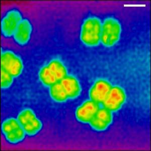
Ultra-high resolution imaging, using X-rays, is leading to significant advances in the field of microscopy in imaging nanostructures of biological samples. The scientists produced the first images of biological cells using the technique

An ultra-high resolution imaging technique, using X-rays, leads to significant progress in the field of microscopy in imaging nanostructures of biological samples. In the journal Proceedings of the National Academy of Sciences, scientists report progress in implementing the "lensless" X-ray microscope approach.
The scientists produced the first images of biological cells using the technique.
A more successful ability to see the cell structures at nanometer scales can yield an important vantage point on the way to the biological and biotechnological revolution. In the case of the bacterium D.Radiodurans for example, this can help settle the question of whether the DNA structure of this organism has nuclear regions that contribute to its resistance to ionizing radiation. By demonstrating the technique, the scientists showed how the biological cell sample can be laid out in XNUMXD imaging.
X-ray imaging is particularly well known in medical applications, such as the CT machine. Despite this, the use of X-ray allows much more than normal imaging. Especially the X-ray radiation with a short wavelength that allows receiving different types of microscopy capable of reaching nanometer resolution. One of the obstacles in achieving high resolution in X-ray microscopy is the difficulty of creating high-quality X-ray lenses. In order to overcome the obstacle, "lensless" microscopy methods were founded in the last decade. This is a technique developed by scientists in the biomedical group of the TUM University in Munich. The technique has raised hopes for ultra-high resolution imaging of materials and biological samples.
The imaging technique, known as ptichography, was first introduced in the 70s. It continued to develop in the field of refractive pattern measurements in which a sample is illuminated with weak light. While the use of this technique in electron microscopy is limited, pythichography has gained enormous popularity among the X-ray imaging community in recent years, thanks to the development of Franz Pfeiffer, who now heads the Biomedical Physics Group at TUM. A critical step in the development of haptychography was published by the team a year ago.
Now, a collaboration between Pfeiffer's group and scientists at the University of Gottingen in Switzerland has advanced the initial development of imaging biological cells.
The results show that lensless X-ray imaging, especially pythichography, can be used to accurately map the electron density present in a biological sample. Biological samples are very delicate and almost invisible to X-rays, so creating such a precise measurement technology is a challenge in itself.
Pfeiffer's group is looking ahead and working on further improvements in the technique. In particular, his team intends to focus on XNUMXD imaging of biological samples.

10 תגובות
What is not clear? Bacteria were seen a long time ago (Lewenhoek, 1673) but with the new method you can see structures inside the bacteria, for example localization and structure of the DNA and I assume also large complexes. There are also other methods for the purpose, but apparently with the current method they were able to see intracellular structures that could not be seen before.
Another thing that may be trying to achieve here is live imaging compared to an electron microscope which requires the killing of the cells and does not allow to look ON line at processes.
By the way, the bacterium in the picture Deinococcus-radiodorans is a very interesting bacterium, all four pseudo-cells that are visible are actually one bacterium that contains four copies of the genome and constitutes one cell. As mentioned, this bacterium has an extraordinary resistance to radiation that has been extensively studied at the Weizmann Institute by Prof. Avi Minsky.
Do not understand. X-rays are used to know the structure of molecules according to their interference pattern (see the ribosomes story), but only now can you see bacteria?
Someone explain the contradiction.
No. Bacteria and molecules are not of quantum size and usually do not exhibit quantum properties.
According to the quants, subatomic particles have no definite location either in space or time, and they are in infinite places all the time, so if this is true how can you see them? , after all, they are supposed to come out blurred in the image, and smeared, and it doesn't matter what type of microscope we use.
Is it at the level of photons and electrons what you see in the sample?
Usually this is not important when looking at cells and organic substances like this. For example, in confocal microscopes (which are popular in the field of biology) the films you make are like taking many individual pictures one after the other.
The link you sent is very interesting. According to my understanding, they send the X-ray beam that passes through the sample and creates a diffraction pattern behind it. With the help of mathematical adjustments, it is possible to understand the size and depth of the sample at any given place - this is how you discover the internal structure and density of the material in the small scales inside the bacteria.
The degree of accuracy in the method depends only on the wavelength of the projected light, which is probably why X-ray is good for the method.
A PDF link that explains the technique "Ptychography" is in English and quite complicated to understand (at least for me) so whoever wants:
https://documents.epfl.ch/users/f/fp/fpfeiffe/www/pr/dierolf_epn_2008.pdf
Were these telescope shots taken in "real time"? Or is it individual pictures?
The bacteria are micro in size, but the cell structures inside them are on the nano scale. In the picture you can see the areas inside the bacteria where the density is highest, colored red. The low density areas are colored blue.
The image shows slightly blurred bacteria. Their actual size is around the order of a micron. Where does the nano element come in here?