Soon we will be able to obtain extremely detailed images of the molecules of life at atomic resolution, thanks to the overcoming of the phenomenon of destruction of biological materials due to the mere observation of them, by three scientists who were recognized already in the first stages of using the device in laboratories
Soon we will be able to obtain extremely detailed pictures of the molecules of life at atomic resolution. The Nobel Prize in Chemistry for 2017 is awarded to three researchers: Joachim Frank from the USA, Jacques Dubusche from Schoenitz and Richard Henderson from the UK for the development of innovative cryogenic electron microscopy that simultaneously simplifies and improves the imaging of biomolecules - the molecules of life. This method advanced the field of biochemistry into a new and fascinating era.
Image is the basic key to human understanding. Scientific breakthroughs were based in many cases on the successful visualization of objects invisible to the human eye. However, there are still many gaps in biochemical maps due to the fact that the existing technology in this field has difficulty producing clear images of the molecules of life. The method of cryo-electron microscopy changed the situation from end to end. Researchers are now able to freeze the minute movements of biomolecules and observe processes that until now were completely unknown, processes that are essential both for understanding the biochemistry of life and for the development of more effective and advanced drugs.
For a long time, researchers believed that electron microscopes were only suitable for viewing dead material, because the powerful electron beam destroys any living biological material. However, in 1990 researcher Richard Henderson was able to use an electron microscope to obtain a XNUMXD image of a protein at its atomic level. This breakthrough demonstrated the tremendous potential of this method.
The researcher Joachim Frank made the method more common and simpler. Between the years 1975 and 1986 he developed a method for image processing during which the two-dimensional and ambiguous images obtained from an electron microscope are analyzed and merged while revealing the three-dimensional structure in a clear and sharp way.
Jacques Dubois added water to the electron microscope. Liquid water evaporates in the vacuum chamber used in electron microscopes, so the biomolecules break up. In the early XNUMXs Dubusha succeeded in vitrifying water - he cooled the water at such a high speed that it became a glassy solid around the biological sample, thus allowing the biomolecules to remain in their original form and not break down, even in a vacuum chamber.
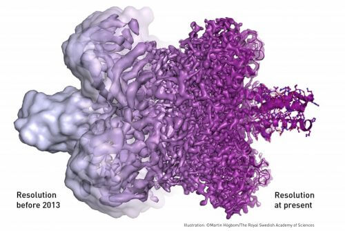
Following the contributions of these three researchers, the field of electron microscopy has improved tremendously. In 2013, the researchers succeeded in achieving the required atomic resolution, and today biochemistry researchers are able to produce 2017D structures of biomolecules routinely and daily. In the years since then, the scientific literature has become replete with wondrous images of countless biological molecules, from proteins responsible for antibiotic resistance to the detailed structure of the Zika virus. Thanks to the contribution of the three winners of the Nobel Prize in Chemistry for XNUMX, the field of biochemistry is now standing at the threshold of a new and fascinating era whose era is just beginning.
