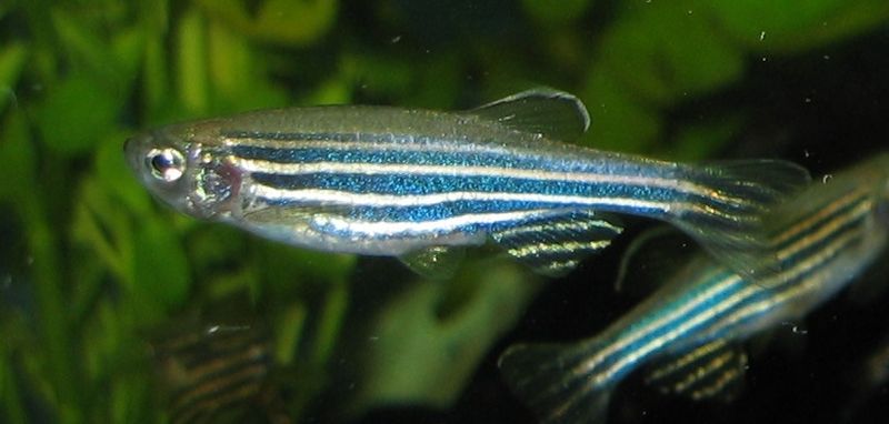
The research may shed light on medical problems such as the way cancer cells spread, why some people are more exposed to heart attacks or what role genes play in addictions

Mayo Clinic researchers have designed a new tool that helps discover the function of proteins based on the genetic code. The team of researchers, headed by Dr. Steven Acker, was able to stop the operation of separate genes and then resume their operation and observe the development of the embryos and fry. The study was published in the journal Nature Methods.
The research may shed light on medical problems such as the way cancer cells spread, why certain people are more exposed to heart attacks or what role genes play in addictions. With this method, it is also possible to investigate more complex issues, such as the genetics of behavior, the flexibility and memory of cells, stress, learning and epigenetics.
The study conducted at the zebrafish facility (Zebrafish Core Facility) of the Mayo Clinic may contribute to the unification of biology and genetics and describe the complex fabric of the interrelationships between DNA, gene function, and the expression or transition of genes and proteins. In the study, the protein expressions and function of 350 locations (loci) among about 25,000 protein-coding genes were examined. The researchers intend to discover 2,000 more places.
"To me, this system is a toolbox that can provide answers to fundamental scientific questions," said Dr. Acker, a Mayo Clinic molecular biologist and lead author of the paper. "The results open the way to a biological field that until now was impossible, or impractical, with the existing methods of genetic research."
For the first time ever…
The research includes several pioneering achievements in the study of genetics. These achievements include a reversible ability to efficiently insert a transposon mutagen. In almost all the places examined, the rate of endogenous expression exceeded 99 percent.
The study yielded the first collection of non-mouse conditional mutant alleles; In contrast to the popular conditional alleles in mice, which switch from active to inactive, the conditional mutants in zebrafish switch from inactive to active - thus offering new insights into the local requirements of genes. The transposon system includes mutant chromosomes with fluorescent labeling, which opens the door to a new genetic world that until now was very difficult to achieve, or could not be achieved at all, with the conventional mutagenetic methods, such as chemical or retroviral insertion.
The project also represents another first in vertebrate mutation. Thanks to the natural transparency of the zebrafish's body, the researchers were able to record the function of the genes and the dynamics of the proteins, and find connections between the expression of the genes or proteins and the function in a single system, to provide direct genetic links.
The researchers intend to integrate information from the research into a new genetic codex, which can be used as a reference point for the information stored in the genome of vertebrates.
Shed light on diseases...
The researchers revealed that zebrafish are transparent to transposons - also known as jumping genes - because they move throughout the cell's genome. The transposons instructed zebrafish cells to mark mutant proteins with a fluorescent protein 'tag'.
"This way you can investigate a whole new set of problems" said Dr. Acker. "This adds another level of complexity to the genome project."
Dr. Acker's team breeds about 50,000 zebrafish in the Zebrafish Core Facility. To observe mutations, photograph and document them, in the numerous and tiny fish, team members collaborate with researchers from around the world and also assist teachers from Rochester Public Elementary School. As part of the Mayo Clinic and Winona State University program, which is called InSciEd Out (Integrated Science Education Outreach), the teachers document mutations and learn about the scientific method.
The other members of the research team include Dr. Carl Clark; Dr. Yong Ding; Stephanie Westcott; Victoria Badel; Tammy Greenwood; Urban soup; Kimberly Schoster; Dr. Andrew Petzold; Dr. John Nee; and Dr. Kasialoi Xo, all from Mayo Clinic; Dr. Darius Blackiones; Audrey Nielsen; and Dr. Sridhar Sivasobu, all from the University of Minnesota; Dr. Hans-Martin Puguda, and Dr. Matthias Hermschmidt from the University of Cologne in Germany; Vashok Pathovari and Dr. Vinod Sakaria from the Institute of Genetics and Integrative Biology in New Delhi.
The study was funded by the National Institute on Drug Abuse, the National Institute of General Medical Sciences, the National Institute of Diabetes and Digestive and Kidney Diseases, and Mayo Clinic.

2 תגובות
transposons.
The article lacks a lot of basic information.
What is the technique used in connection with transpososomes? How is it different from everything that has been done to date?
How is it different from any other 'scanning' method that has been done to date that has been published in
Nature Methods?