A new model explains the action of the ultrasonic wave on the living cell

A new model developed at the Faculty of Biomedical Engineering explains the action of ultrasound waves on the living cell. The model, which was presented in PNAS (Journal of the American Academy of Sciences), was developed last year by Professor Eitan Kimmel, Professor Shay Shoham and Dr. Boris Krasovitsky.
The model explains phenomena observed years ago in goldfish, using electron microscopy, in Dr. Viktor Frankl's doctoral thesis under the guidance of Professor Kimmel.
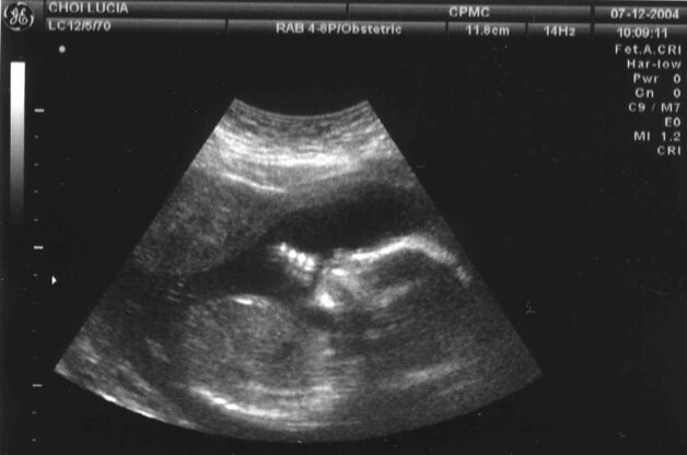
Ultrasound is a popular and important imaging technology, used among other things in medicine. Since it is a non-invasive technology that can be used in a targeted manner, the use of ultrasound is also increasing for treatment and healing purposes. However, the mode of operation of the ultrasonic wave in these uses, and the interaction between it and the living cell, were not clear until now.
"Our model combines the physics and dynamics of bubbles with the biomechanics of the cell," says Professor Kimmel. "The model basically explains the origin of the bubbles that form in the body's tissues under the influence of ultrasound."
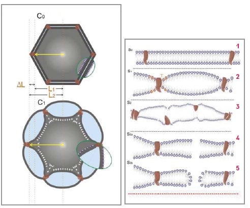
According to the aforementioned model, the ultrasonic wave exerts mechanical energy on the cell membrane (membrane), which is made of two monomolecular layers of fat. This energy causes the intra-membrane space to swell and contract. "This is the essence of the interaction between ultrasound and biological tissue, and this is the source of the bubbles that form in the tissue," says Professor Kimmel.

diverse applications
The Technion researchers estimate that the new model will enable the development of different and diverse applications such as the introduction of drugs into the brain and tumors in the body's tissues, modulation (proactive activation of areas of the nervous system, for example among paralyzed people), pain suppression, encouraging the growth of blood vessels and the fusion of wounds and bones. However, they emphasize, it is important to redefine the safe intensity ranges in the use of ultrasound. "The operation of the ultrasound affects the function of the cell proteins and the permeability of the membrane. At certain intensities this may have positive consequences for the cell, but higher intensities may cause it damage - as you can see in the lower diagram on the right. That is why we recommend continuing to investigate the issue according to the model we have developed."

2 תגובות
Does the transducer create a single wave with one defined frequency, or a continuous frequency domain or single frequencies (lines)?
An ultrasound bath makes a noise. Is the noise in the audio field created by the ultrasonic transducer?
How is the power distributed between the different frequencies?
Is there a TLV - for ultrasonic radiation?
Which organ in the body is most sensitive to injury from exposure to an ultrasonic wave?
How do you protect yourself? Is there a protector such as glasses, earplugs?
Thanks
Now everyone will start to be afraid to use ultrasound