There is no doubt that radiation at high levels is a factor that is very harmful to health and causes various diseases, but the hormesis model claims that the same cannot be said about radiation at low levels, the amount of radiation given in various imaging tests

Ionizing radiation is considered by doctors and subjects to be avoided as much as possible. When subjects are referred for testing, they fear the radiation they are going to receive. When people consider studying radiology or specializing in radiology, the exposure to radiation can alert them and be a consideration in their decision. There is no doubt that radiation at high levels is a factor that is very harmful to health and causes various diseases, but can the same be said about radiation at low levels, the amount of radiation given in various imaging tests? Not sure.
In January 2017 it was published Article in the journal Journal of Nuclear Medicine) JNM) in which the researchers claim that exposure to medical radiation does not increase a person's risk of getting cancer. The authors claim that the common belief that low doses of radiation, such as those obtained in medical imaging, increase the risk of cancer, is incorrect and that they are based on an old hypothesis, 70 years old, that has not been sufficiently investigated.
In December 2016 it was published A cohort study (a large observational study done over time) in the journal Radiology in which researchers compared the cancer incidence and mortality rates between 43,763 radiologists and 64,990 psychiatrists who graduated from medical school between 1916 and 2006 (psychiatrists were chosen as a comparison group because they are not likely to be exposed to occupational radiation) . In the study, it was found that radiologists who graduated in the last decades, in which there are accessories for radiation protection, not only did not tend to get cancer more than the group of psychiatrists, but were also healthier and had a lower risk of cancer and heart disease. We will present these two studies in a broader way later on.
Today's research data regarding radiation is mainly based on a study done on the survivors of the atomic bombings (LSS study; Life Span Study), but in this study quite a few methodological mistakes were discovered. Article, which was published in December 2018 in the journal Genes and Environment and reviewed this study, claims that the study's control group was flawed because it was also exposed to secondary radiation and that the facts known today (and which were not known when the LSS study was done) are that the average life expectancy of atomic bomb survivors is Longer than the average lifespan of other Japanese who were not exposed to this radiation. Deaths as a result of a solid cancerous tumor of the survivors of the atomic bombs and of the control group (which, as mentioned, was not suitable because it was also exposed to radiation) are also low compared to the Japanese average. This study will also be discussed later in the article in a more extensive manner.
The debate among scientists in the field of low-dose radiation is expressed in three radiation models that exist today: the linear model without a threshold, the threshold model and the hormesis model of radiation. This article presents studies related to these models - strengthening or weakening them. Later we will focus on the hormesis model of radiation and review several epidemiological studies related to radiologists/radiologists/other doctors who are exposed to radiation and what are the health consequences of this exposure. References to studies and references are integrated within the article and in the bibliography at the end of the article.
It is important to note that the model currently accepted in the world by all medical associations is the linear model without a threshold and the role of this article is not to discredit the model but to provide information in the context of studies that support it versus studies that support the hormesis model.
Radiation exposure models
Ionizing radiation is the radiation of particles or electromagnetic waves with high energy capable of ionizing a substance, that is, releasing electrons from atoms or molecules. The ionization ability of a particle or photon depends only on its energy and not on the number of particles. That is, even a large amount of particles or photons with low energy still constitute non-ionizing radiation.
We constantly absorb ionizing radiation from nature. This radiation comes from four main sources: radiation from radon gas (the main cause of ionizing radiation), cosmic radiation, solar radiation and radiation from natural sources (referring to radiation from materials containing radioactive atoms, which are found, for example, in the walls and floors of the house, in the soil and rocks outside the house).
Radon gas, as mentioned, is the main cause of ionizing radiation. It is found in uranium-containing rocks, is released by diffusion, and accumulates in unventilated places such as basements of houses. The intensity of this radiation varies greatly from place to place and some speculate that Hadron gas is the second most important cause of lung cancer in the United States.
So how does the world of science treat the effect of radiation on us? In general, we can say that today there are three models in the field of radiation exposure:
The first model is The linear model without a threshold (linear no-threshold; LNT).
The model was introduced in 1956 by the United States Academy of Sciences (NAS). As mentioned, this is the model that currently dominates the world of science. He claims that ionizing radiation is always harmful, regardless of its dose, and that the sum of several very small exposures is considered to have the same effect as one larger exposure (the linearity of the response).
The second model is The threshold model (threshold theory), which assumes that exposure to low levels of radiation is not harmful. This model represents the scientists who disagree on the LNT theory regarding the damage of low dose radiation.
The third model is called The radiation hormesis model (radiation hormesis). This model, which in this article we will review studies related to it, claims that exposure to radiation at very low levels, such as the radiation levels used in medical imaging, can even have a protective effect on the people exposed to it.
The ongoing controversy that divides these three theories is the effects of low amounts of radiation on human health. While the effects of large, immediate amounts of ionizing radiation are understood and easily seen in humans (eg, Japanese atomic bomb survivors), the effects of low-level radiation are very difficult to observe and these effects are controversial. The reason for this is that when the basic cancer rate is already very high, and the risk of developing cancer reaches 40% due to individual lifestyle and environmental influences, it is not clear how much the effects of exposure to low-level ionizing radiation contribute to this. The greatest controversy in this field exists regarding the hormesis model of radiation. Can radiation at low levels not only not harm, but even benefit us? We will deal with this in the next section.
The hormesis model of ionizing radiation
Hormesis (in English hormesis) is a term that originates in the world of toxicology (a field that deals with the study of the negative effect of chemicals on living beings). This term indicates a phenomenon in which a small amount of poison or toxin actually has a beneficial biological effect. The biochemical mechanisms that cause the phenomenon are not fully understood, but it can be assumed that the toxin triggers the activation of defense and repair mechanisms, which succeed in overcoming the negative effect, and even immunize the body against future toxin attacks. When we talk about hormesis, we are not talking only about toxins, but about any activity that, in the right amount, can strengthen the body's defenses. Possible examples of hormesis are for example:
- Although moderate physical activity causes the body to go into a state of stress, it nevertheless has a beneficial effect on health.
- In animal experiments it was found that calorie restriction, a certain state of stress, extended their lifespan.
- Several studies have shown that consuming a small amount of alcohol per day may contribute to the prevention of stroke and heart disease.
- Mitohormesis - the mitochondria in the cells create free radicals that cause the cell to activate antioxidant mechanisms that also protect against free radicals that come from outside the cell.
- The hygiene hypothesis claims that exposure to pollutants at a young age contributes to the proper development of the immune system.
The hormesis theory of ionizing radiation claims that low levels of ionizing radiation are beneficial because they stimulate the activation of cellular repair mechanisms that protect against disease (repair mechanisms that might not have been activated if there had been no exposure to ionizing radiation). The increased ability of the repair mechanisms, as the theory claims, not only later helps the body deal better with ionizing radiation, but also inhibits diseases that are not related to radiation exposure.
What is meant by low radiation levels?
When referring to measuring radiation, to its absorption in the body (because what is irradiated to the body is not equal to what is absorbed by the body), there are three types of units:
The first unit is the absorption unit Gray (Gray) or for short (Gy). One gram is equivalent to one joule of energy absorbed by one kilogram of body mass.
The damage caused to the cells is measured in units of Sievert or for short (Sv). A radiation dose of one gram of beta, gamma or X-ray radiation causes one Sievert of damage. One Geri dose of alpha radiation causes 20 Sievert damage. The Sievert is a rather large unit, so it is customary to work with millisieverts (mSv) or microsieverts. In the United States and also in Israel, it is still customary to use the unit called rem (rem) or millirem (mrem) instead of Sievert (one Sievert is equivalent to 100 Rem) and in the past they also used Rad instead of Gray (one Gray is equivalent to 100 Rad). Radiation levels can be detected and measured using a Geiger counter or dosimeter.
How much radiation do we receive during our lives?
Of course, these are variable values related to the location and human activity. As mentioned, some of the radiation we are exposed to is natural radiation. The natural radiation levels, as a result of the modern lifestyle, are below 4 millisilvert cumulative radiation with a standard deviation of 1 millisilvert (this is the average terrestrial and cosmic radiation, with the exception of the radiation emitted from the radon gas, which mainly affects the lungs). This amount of radiation should not cause problems for the average person, on the other hand There are studies which show that children who are particularly sensitive to radioactivity, even at natural levels of radiation, develop leukemia and other types of cancer. there is also Studies who claim that exposure to high natural radiation causes neurological damage, such as low achievements of students in Sweden in areas with high natural radiation. Many flights also increase the amount of exposure to radiation - for example, a 14-hour flight from New York to Tokyo exposes the passengers to 0.1 millisilvert.
Medical imaging is also a component of the total amount of radiation that a person receives during his lifetime. The values vary, but in general, in a chest photograph, the subject receives up to 0.06 millisilvart. The effective dose in a typical abdominal CT scan is about 10 millisiliverts (the values are lower in radiation-reduced scanners) - these are all low levels of radiation.
Are these radiation levels dangerous for the average person?
The research literature claims that an acute dose of 100 millisilvart can increase the risk of cancer by 0.8% - there is almost no dispute about this. The dispute is what happens at lower radiation levels, radiation levels that are given in the cases mentioned above. Are they harmful, ineffective or beneficial to the person exposed to them. The hormesis model claims that they are beneficial to the health of the exposed person. He claims that exposure to radiation, similar to or slightly above the natural background radiation, is not only not harmful, but even beneficial in a certain location.
The controversy regarding the hormesis model of radiation - a review of studies
The supporters of radiation hormesis claim that the repair mechanisms within the cells and in the immune system that are activated during exposure to ionizing radiation, not only protect against the destructive effects of radiation but also work to eradicate cancer cells that are formed spontaneously, regardless of exposure to ionizing radiation.
Low doses of radiation are considered levels below 100 millisilverts and a radiation exposure rate is considered low when it is below 0.1 millisilverts for less than a minute. While epidemiological studies of populations of people exposed to acute and large amounts of ionizing radiation, such as the survivors of the atomic bombings of Japan, have strengthened the LNT model, studies involving low levels of radiation and low levels of radiation over time have failed to demonstrate an increase in cancer rates.
The reason for this is, as already mentioned above, that at the very least the basic cancer rates are very high (42 people out of 100 people will be diagnosed with cancer in their entire life) and the chances of getting cancer (about 40%) vary due to lifestyle and other environmental influences - all of this It makes it difficult to distinguish the subtle classes, which may be caused, following exposure to ionizing radiation in a low amount. Epidemiological studies can detect an increase in cancer rates of about 1.2 to 1.3 height, meaning about a 20% to 30% increase in risk, but for low radiation doses (between 1 and 100 millisiliverts), the predicted increase in cancer incidence rates is 1.001 to 1.04 and a number The occurrence of cancer cases, if they exist, cannot be detected due to misleading variables, sampling errors, etc. For example, variables of smoking prevalence, and even inaccuracy in reporting about smoking, can cause such errors and measurement problems. Thus, even a study with a large number of subjects, some of whom did not accurately report their smoking habits, would not be able to detect the effects of low levels of radiation any more than a smaller study that would compensate for the prevalence of smoking. When there is really no epidemiological evidence, there is much debate about what the dose-response relationships of ionizing radiation below 100 millisilvart really are.
The strongest evidence to support the hormesis model comes precisely from the field of laboratory research. Cell culture studies can be useful in order to find mechanisms behind biological processes, but they can also be criticized for not being able to effectively express the activity of the living organism.
study done by EI Azzam suggests that early exposure to radiation causes cells to activate their defense mechanisms. Another study done by de Toledo and his associates, showed that irradiation with radioactive gamma rays increases the concentration of glutathione, an antioxidant found in cells. in 2011, in vitro study led by SV Costes showed in images a strong non-linear reaction sequence of certain repair mechanisms in the cell which are called radiation-induced foci (RIF). The study found that a low dose of radiation caused more formation of RIF than high doses of radiation and that even after exposure to a low dose of radiation, the RIF continued to form (at 2 Gray, 15 RIF/Gy were formed while at 1 Gray, 64 RIF were formed /Gy). These results suggest that low amounts of ionizing radiation do not increase the risk of cancer proportionally to the dose of radiation and therefore constitute a contradiction to the LNT model (linear-no-threshold standard model).
Mina Bissell, a global researcher in the field of breast cancer and a partner in the research mentioned She declared: "Our data show that at low levels of ionizing radiation, the DNA repair mechanisms work much better than when the cells are irradiated with high levels of radiation. This non-linear response to DNA damage following radiation casts doubt on the general assumption that any amount of radiation is harmful."
It is important to note that on the other hand there are laboratory studies that support the linear model without a threshold. Studies on particles In laboratories it is demonstrated that the passage of even one alpha particle (for example an alpha particle originating from radon gas) through a cell nucleus is highly mutagenic (meaning it causes mutations) and that when it comes to alpha radiation, The risk of mutations is higher at low doses (even if very few cells were damaged by alpha particles). This confirms what is suggested by the linear model without a threshold, however, Currently there is not enough evidence In order to suggest that this effect promotes the formation of cancer cells in humans at low doses.
In addition to laboratory studies, were also carried out Animal studies In order to confirm or deny the hormesis model:
A relatively old study (from 1955) on mice exposed daily to low levels of radiation suggests that those mice can live longer compared to mice that did not receive such radiation (the control group). Further research , which was done on mice led by Miyachi, found that exposure of the mice to a dose of 200 mGy X-ray protected the mice both against further future exposure to radiation, and also against ozone gas (a gas that filters ultraviolet radiation from the sun in the earth's sestosphere but is toxic on the earth's surface- causes breathing difficulties and various irritations).
In another study On rodents, a researcher named Sakai and his associates found that 1 mGy/hr (1 micro gray per hour) of gamma radiation prevents the formation of cancer in the rodents (cancer created by injecting a substance called methylcholanthrene, which causes cancer). In an article published in 2006, a dose of 1 Gy of gamma radiation was irradiated into cells (at a constant rate from a radioactive source) over a long period of time. Also in this study it was proven that low levels of radiation protect the cells in the future more than high levels of radiation. Early exposure to gamma rays increased the cells' ability to cope and prevent tumor changes when a second dose of radiation was given. A similar study On dogs it showed that there are no more cancer cases nor a decrease in the life expectancy of dogs that were irradiated with 3 mGy per day.
BIR Report V He referred to the results of these studies and argued that the interpretation of these results should be treated with caution, because these doses can also increase the risk of certain types of cancer. The authors of the report agreed that low-level radiation exposure can be interpreted as an adaptive effect at the cellular level, but it should also be noted that the exposed animal population experienced a high incidence of myeloid leukemia and other myeloproliferative disorders. Below is a quote from the report in this regard:
"The acquisition of the ability to protect against radiation (radio resistance) and the ability to repair other cells under selective selection and the mutagenic pressure of long-term radiation administration (chronic radiation) are temporally and causally related to the formation of leukemia ((leukemogenic following exposure to radiation).
is also Human studies were carried out in the context of the hormesis model.
Central research who presented a phenomenon of radiation hormesis and tested the chances of lung cancer among those exposed to radon gas. This gas is a significant source of radiation for humans and it is usually recommended that its concentration be less than 150 Bq/m³ (Bq per cubic meter). A retrospective study of lung cancer odds showed a significant decrease in cancer rates in a group exposed to 50 to 123 Bq per cubic meter of radon gas, compared to a group exposed to 0 to 25 Bq per cubic meter of radon gas. It is important to note that other studies that examined home exposure to radon gas did not report hormetic effects in their results (including Extensive research called the "Iowa Radon Lung Cancer Study" done by Field et al in 2000).
Another study related to the hormesis model is study that tested exposed to a very high rate of natural gamma radiation in the city of Kerala in India and the cancer rates. The sand of Kerala contains a reddish-brown phosphoric mineral called monazite which is made up of several elements. Monazite may be radioactive, as it contains thorium and the sands of Kerala contain one-third of the world's radioactive reserves of radioactive thorium. This sand emits about 8 microsieverts per hour of gamma radiation, 80 times the emission in London, but study Conducted over a decade on 69,985 local residents, and published in the journal Health Physics, did not find an increased risk of cancer in them, which rules out a statistically significant positive or negative relationship between background radiation levels and the risk of cancer in this sample, and basically opposes the linear model of radiation exposure.
In an Australian study, who analyzed the relationship between exposure to ultraviolet solar radiation (UV) and DNA damage, the results showed that although the frequency of cells with broken chromosomes increased with increased exposure to the sun, the lack of repair (misrepair) of the DNA strands decreased as the solar radiation increased ( meaning the repair capabilities of the cells have increased).
The health of residents of radioactive buildings in Taiwan received prominent attention in the popular Hormesis treatments. In 1982, more than 20,000 tons of steel accidentally arrived contaminated with cobalt 60. Much of this radioactive steel was used to build apartments and thus thousands of Taiwanese residents were exposed to reductive radiation levels in the form of gamma radiation 1000 times higher than normal background radiation (an average of 47.7 mSv, maximum 2360 mSv excess cumulative dose). It was only in 1992, ten years after the houses were built from radioactive steel, that the omission was discovered.
Medical research published in 2004 claimed that cancer death rates in the exposed population were much lower than expected. However, this initial study did not control for the age of those exposed and compared a much younger population (average age of 17.2 years at initial exposure) with the much older general population of Taiwan (average age of about 34 years in 2004) - this is a very serious research flaw , as it is known that older people have a much higher chance of cancer even in the absence of radiation exposure.
A follow-up study by Hwang et al In 2006 he found that the incidence of "all types of cancer" in the irradiated population was 40% lower than expected (95 cases instead of 160.3 expected cases), except for leukemia in men only which was relatively higher than expected (6 cases versus 1.8 expected cases) and except for thyroid cancer which in women which was higher than expected (6 cases versus 2.8 expected cases) - where this increase in the number of cases was found only in those who were exposed before the age of 30.
Hwang and his partners suggested that the explanation for the results of this study is that a lower rate of "all types of cancer" could be due to the fact that the exposed population was of a higher socioeconomic status and therefore had a healthier lifestyle and therefore got less sick - but it was difficult to prove this. In addition, they warned that leukemia was the first type of cancer to develop in survivors of the Hiroshima and Nagasaki bombings, so it could be decades before tumors were detected in the more common types of cancer.
Besides the excess risks of leukemia and thyroid cancer, Posting later In the context of this study, he noted several different DNA abnormalities (e.g. chromosomal translocations) and other health effects among the exposed population (suppression of peripheral neutrophilic leukocytes, increase in eosinophils, changes in lymphocyte subpopulation distributions, delays in physical development among exposed children, increased incidence of lens opacities The eye - cataracts, increased risk of abnormalities in the thyroid gland, changes in the hematopoietic cells - stem cell source of the blood cells - in children).
Another layer to resolve the controversy in the field of radiation models was proposed precisely from the direction of Effect of lack of radiation About the organisms living on the surface of the earth and adapted to the level of radiation here.
A panel of experts that met in 2006 in Carlsbad, New Mexico, proposed the establishment of an ultra-low-level radiation laboratory.
It is important to remember in connection with this, that life on Earth developed under low levels of radiation and therefore it is very important to know what the consequences of an "almost no radiation" situation are. The laboratory, if built, will investigate the effects of an "almost radiation-free" state on laboratory animals and cell cultures, and will compare these groups to a control group that was exposed to natural levels of radiation. For the groups that "will hardly be exposed to radiation", potassium 40 (a radioactive substance that contributes greatly to the natural radioactive radiation that we are all exposed to) will be removed from their food. The team of experts believes that the laboratory for ultra-low radiation is the only experiment that can correctly investigate the effects of low-level radiation and then confirm or cancel the assumptions made by the various theories of radiation such as the LNT theory, the threshold theory and the hormesis theory of radiation.
First results of the effects of "almost no radiation" on cell cultures were already reported by two research groups in 2011 and 2012. Researchers from the United States They studied cell cultures, which were isolated in a steel chamber 650 meters underground in an isolated waste plant in Carlsbad, New Mexico, while researchers in Europe reported the effects of "almost no radiation" on mouse cells. Their results are still very preliminary.
Statements of leading nuclear bodies
The radiation hormesis theory has not been accepted by the United States National Research Council or the National Council on Radiation Protection and Measurement (NCRP). In May 2018, the NCRP published Report of an interdisciplinary expert group who critically reviewed 29 qualitative epidemiological studies on populations exposed to low-dose radiation at a low exposure rate, published mainly in the last ten years. The expert group concluded following this review that the latest epidemiological studies support the continued use of the LNT model for radiation protection. This conclusion is consistent with rulings of other national and international scientific committees, based on older data. They found no other model, which explains better than the LNT model, the exposure to radiation and its consequences.
The UN Scientific Committee on the Effects of Atomic Radiation (UNSCEAR) also wrote in its latest report:
"Until the lack of clarity regarding exposure to radiation at low levels is resolved, the committee believes that the risk of cancer is proportional to the amount of radiation, according to the amount of knowledge accumulated to date. Despite this, a straight linear line is not a reaction that should be expected in all situations".
The idea of radiation hormesis was also rejected by the National Research Council's, which is part of the National Academy of Sciences following 16 years of long-term research into the biological effects following exposure to ionizing radiation: "The scientific research database demonstrated that there is no exposure threshold at which radiation can be determined Ionization can be harmful or beneficial. The health risks - especially the development of solid cancer in the organs - increase proportionally with the exposure," he said Richard R. Munson said, Associate Dean of Professional Education and Professor of Epidemiology, Harvard University School of Public Health, Boston.
Later the same committee claimed: "The possibility that a low dose of radiation may have beneficial effects (a phenomenon often referred to as "hormesis") has been the subject of much discussion. Evidence for hormetic effects is reviewed, emphasizing studies published since the 1990 BEIR V study on the health effects of exposure to low levels of ionizing radiation. Despite examples of radiation protection phenomena that can be found at the cellular level or in animal biology, the amount of experimental information available in the field does not support the claim that low levels of ionizing radiation have a beneficial effect. The mechanism of any such possible effect remains obscure. At this stage, the assumption of the hormesis model, which claims that the health benefit to humans as a result of exposure to low levels of ionizing radiation exceeds the possible negative effects from exposure to radiation of such intensity, is not justified."
Although most major consensus reports and government bodies currently adhere to the LNT model, The report of the French Academy of Medical Sciences (French Academy of Sciences-National Academy of Medicine), on the effect of low-level radiation, rejected LNT as a scientific model of cancer risk at low doses. Their claim was that more studies are needed on low radiation doses (below 100 millisilvers and also under 10 millisilvers), taking into account the age of the person and the type of tissues irradiated. Despite the convenience of working with a linear model for regulatory purposes of monitoring radiation exposure, they note that 40% of laboratory studies on cell cultures and animals indicate some level of chemical or radiobiological hormesis.
Do radiologists/radiographers/people who are exposed to radiation get sicker than others?
After reviewing the studies related to the possibility of the hormosis model being correct or refuting it, we may find the answer in the studies that were done on people who are constantly exposed to low-dose radiation as a result of their work - such as radiologists, radiographers, doctors and other people.
The relationship between exposure to radiation and the development of cancer is mainly based on populations that were exposed to relatively high amounts of ionizing radiation (for example: the survivors of the atomic bombs dropped on Japan). The types of cancer associated with large amounts of radiation are leukemia, breast cancer, liver cancer, lung cancer and more, while the period between the exposure to radiation and the discovery of the cancer is known as the "dormant period".
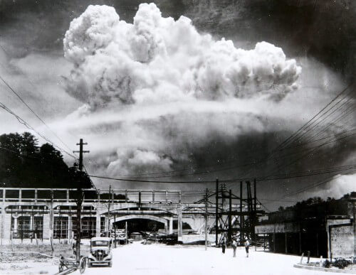
A high amount of radiation kills cells, while a low amount causes damage or even changes in the genetic code (DNA) of the irradiated cells. Large amounts of radiation can kill such an amount of cells that tissues and organs are immediately damaged. As a result, an acute reaction of the body called acute radiation syndrome is caused. At high levels of radiation, there is a direct relationship between the amount of radiation and the speed with which the signs of radiation will appear and the risk of death. This syndrome was diagnosed in many of the survivors of the 1945 bombings and the rescue workers who dealt with the accident at the nuclear reactor in Chernobyl (134 reactor workers and firefighters were exposed to high doses of radiation and suffered from acute radiation syndrome, of which 28 died).
As mentioned, in this section we focus on people who are exposed on a daily basis to relatively low levels of radiation. There are several studies that summarize results regarding morbidity changes in radiologists, radiographers and other people who are exposed to radiation.
A great first study is a study published in December 2016 in the journal Radiology in which it was found that radiologists who graduated from medical school after 1940 are not at increased risk of death from radiation-related factors such as cancer. The research was carried out by my mother Berrington de González (Berrington de González A), head of the radiation division at the NCI (National Cancer Institute). The study was based on the American Medical Association (AMA) database, a database established in 1906 that includes historical data for more than 1.4 million physicians, residents and medical students in the United States.
In the study, the researchers compared the cancer incidence and mortality rates among 43,763 radiologists and 64,990 psychiatrists who graduated from medical school between 1916 and 2006. Psychiatrists were chosen as a comparison group because they are not expected to be exposed to occupational radiation.
The results of the study showed that male radiologists who graduated after 1940 had a better health profile than their psychiatrist colleagues. The all-cause mortality rate for radiologists was lower and there was no evidence of increased mortality from radiation-related causes, such as cancer or cardiovascular disease.
It is important to note that among radiologists who graduated before 1940, higher mortality rates were found from certain morbidity conditions such as acute myeloid leukemia and myelodysplastic syndrome, which are known to be related to occupational radiation. They also had increased death rates from melanoma and non-Hodgkin's lymphoma. The older radiologists also had a higher risk of cardiovascular disease. The explanation for the difference between the older and younger radiologists is the improvement in radiation protection measures in recent decades.
In another article (which was also briefly mentioned at the beginning of the article), which was published in the January 2017 issue of the "Journal of Nuclear Medicine", the researchers claim that exposure to medical radiation does not increase a person's risk of getting cancer. The authors claim that the common belief that low doses of radiation, such as those obtained in medical imaging, increase the risk of cancer, is incorrect, and that they are based on a 70-year-old incorrect hypothesis.
The research was led by Dr. Jeffrey A. Siegel, president and CEO of Nuclear Physics Enterprises in Marlton, New Jersey. Segal claims that there is currently a policy called ALARA (as low as reasonably achievable), in which they try to give as little radiation as possible in X-ray, CT and nuclear medicine tests, and according to him this policy increases the fear of radiation in doctors and patients - a fear that stems from a lack of information. He claims that the linear no-threshold model (LNT) has not been properly proven by science when it comes to radiation at low levels, especially when certain epidemiological studies even indicate that there is a certain protective benefit in these radiations, in the form of activation and strengthening of the DNA defense mechanisms that were created during evolution. The claim of the researchers is that LNT and ALARA are theories that focus solely on molecular damage while ignoring protective biological responses (focusing on the micro and not the macro). They claim that low doses of radiation induce protective responses and provide enhanced protection against further damage over time, including damage from higher radiation exposure.
Segal continues and claims that the evidence today shows a decrease, not an increase, in the risks of cancer when it comes to screening the doses of radiation that are used in the world of radiology. He refers to the cohort study done on the survivors of the atomic bombs that fell on Japan in World War II (Life Span Study atomic-bomb survivor data; LSS) which showed that the risk of cancer (carcinogenicity) predicted according to the linear model does not work when it comes to radiation doses lower than 200 milligrams (mGy).
The effective level of radiation given in a CT scan is about 10 millisilverts (mSv), in a PET/CT brain scan it is around 5-7 millisilverts and a routine whole body F-18 FDG PET/CT scan is between 12 and 15 millisilverts. Therefore, according to the researchers, there is no reason to avoid or fear radiation when trying to reach the correct diagnosis and there is no reason for diagnoses to be missed due to the fear or avoidance of radiation. The researchers also claim that if the LNT model and its descendant ALARA are defeated, new ways of diagnosis and treatment can open up to medicine. The way to move forward with this is to first of all explain the matter to the doctors and from there this idea will be able to permeate the entire public.
Interesting article On the Scientific American website from 2013, she summarized the available information regarding the question of whether a CT scan, which, as mentioned, is a test that includes a relatively large medical X-ray radiation (equivalent to 150-1100 conventional X-rays or about a year's exposure to natural radiation - depending on the type of test and the scanner), Can cause more cancer cases in the general population?
The article presents studies that claim that approximately 29,000 cancer cases are caused by the 72 million CT scans that were performed in the United States in 2007, and that one cancer case was created for every 400-2000 routine chest x-rays. The point is that those studies are based on data on cancer rates among the survivors of the atomic bombs dropped on Hiroshima and Nagasaki in August 1945 during World War II.
About 25,000 atomic bomb survivors were exposed to a relatively low dose of radiation equivalent to one to three CT scans. Several years after the explosions, researchers began tracking disease and death rates among more than 120,000 survivors. The results showed, for the first time, that the risk of cancer from radiation depends on the dose, and that even very small doses can increase the risk. Based on these data, a report by the National Research Council conducted in the United States in 2006 estimated that exposure to 10 mSv (the estimated dose from a CT scan of the abdomen), increases the lifetime risk of developing fatal cancer by 0.1%. The US FDA reduced this to 0.05%. If we assume that every person in the US has a 20% chance of dying from cancer, a single CT scan increases the average patient's risk of developing a fatal tumor from 20% to 20.05%!?!.
The big problem is that these estimates were based on serious methodological flaws. Among the survivors who were exposed to radiation of 100 millisilverts or less - including the doses typical of CT scans - the number of cancer cases and their deaths is so small that it is almost statistically impossible to be sure that they are significantly higher than the cancer rate in the general population.
To compensate for this, the National Research Council and others based their estimates mainly on data from survivors exposed to radiation levels in the range of 100 mSv to 2 Sv, when they assumed that the cancer risk in relation to the dose is similar when there are high and low radiation levels - but this is not necessarily true. One thing is clear - the number of cancer cases developed in atomic bomb survivors during the rest of their lives is not large enough to provide the statistical power needed to reliably predict the cancer risk associated with CT scans in the general population today.
Two other data that constitute serious methodological problems in these studies:
- The survivors of the atomic bombs received gamma radiation on their entire body, while subjects who perform CT receive radiation mainly on one area of their body - therefore exact comparisons between the two cases are quite problematic.
- The survivors were after a difficult war, had poor nutrition and had a problematic approach to medical treatment - therefore the same level of radiation can cause a more serious illness in those survivors than in healthy people living today.
In conclusion - the data on which the linear radiation model without a threshold is based today can be very problematic, and until there are recent studies on the matter it is difficult to extrapolate from one case to another.
Another article , who found flaws in the research on the survivors of the atomic bombs in Japan and supports the hormesis model, was published in December 2018 in the journal Genes and Environment. In this article the researchers summarize and claim that the model supporting the linear relationship between radiation and damage (the LNT model, the linear model without a threshold) was recommended without solid data by the Academy of Sciences in 1956. In 2006, the Academy gave priority to the BEIR VII report and saw it as support for the LNT model. This report was based on the LSS (Life Span Study) study of the survivors of the atomic bombings. Among the other flaws in the LSS study, the researchers emphasize two main flaws:
- The secondary radiation, to which both the bomb survivors and the control group were exposed, was not taken into account during the study. In particular, the control group was not suitable to be used as a control group.
- The hormesis model is indeed beyond the ability of the LSS study to prove, but the results in the field show that it did occur. The average lifespan of atomic bomb survivors is longer than the average lifespan of other Japanese who were not exposed to this radiation. Deaths as a result of a solid cancerous tumor of the survivors of the atomic bombs and of the control group (which, as mentioned, was not suitable because it was also exposed to radiation) are low compared to the Japanese average.
As a result, the researchers claim that it is reasonable to conclude that the radiation exposure of the survivors of the atomic bombs in Japan extends their life expectancy and reduces cancer deaths on average, which indicates a failure of the LNT model. In their opinion, unfortunately, the LNT model was and is still used today as the model underlying the regulation of radiation exposure and humanity must learn to be less afraid of low-dose radiation and understand that such radiation is not only harmless, it can even be beneficial.
Ionizing radiation and cardiovascular disease
Cardiovascular disease has also been associated with ionizing radiation and several studies have been done on the matter.
First article Worth mentioning in this context is an article from 2017 that deals with cardiovascular morbidity in the context of ionizing radiation. Recent epidemiological studies have revealed that exposure to ionizing radiation not only increases the risk of malignant diseases but also of cardiovascular diseases (and other medical problems such as cataracts in the eyes). In this study, people who survived atomic bombs, or experienced occupational/medical exposure to radiation, were examined for their health. The relationship between the level of radiation and cardiovascular diseases was examined and it was found that the effect of radiation has no threshold, meaning that even with exposure to levels of radiation with an intensity of 1-2 Gray, the risk of cardiovascular diseases increased - although it only became clear 10-20 years after the exposure. The higher the level of exposure to radiation, the greater the risk. On the other hand, due to confounding variables, the researchers have not yet been able to prove that low levels of radiation, below 0.5 Gray, correspond to the linear model without a threshold.
Another article dealing with the issue of low-level radiation and cardiovascular disease is This article, a review article on the field from 2016 - definitely recommended for those who want to get a comprehensive picture of the research in the field.
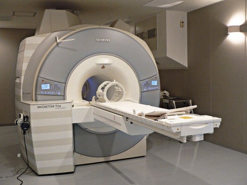
The author of the article: Ofer Ben Horin, who has about 25 years of experience in applications, drug research and training in the field ofMRI and the medical imaging (radiology). Author of the bookMRI the complete guide-medicine and physics meetOn the website www.mriguide.co.il
Bibliography and for further reading
A website explaining radiation and ionizing radiation units:
The effect of natural radiation on leukemia in children in the UK:
The research of Dr. Jeffrey A. Siegel, President and CEO of Nuclear Physics Enterprises in Marlton, New Jersey, which claims flaws in the LHT model:
Jeffry A. Siegel, Charles W. Pennington, Bill Sacks. Subjecting Radiologic Imaging to the Linear No-Threshold Hypothesis: A Non Sequitur of Non-Trivial Proportion. Journal of Nuclear Medicine, 2017; 58 (1): 1 DOI: 10.2967/jnumed.116.180182
An article on the ScienceDaily website about the exaggerated fear of low-dose ionizing radiation:
Society of Nuclear Medicine. (2017, January 9). Fear of diagnostic low-dose radiation exposure is overstated, experts assert. ScienceDaily. Retrieved April 4, 2019 from www.sciencedaily.com/releases/2017/01/170109150038.htm
An article about the survivors of the atomic bombs - an increase in their life expectancy and a decrease in their cancer rates:
An article criticizing the LTH model:
It Is Time to Move beyond the Linear No-Threshold Theory for Low-Dose Radiation Protection
An article dealing with the effects of low-dose ionizing radiation:
Aurengo (2005-03-30). "Dose-effect relationships and estimation of the carcinogenic effects of low doses of ionizing radiation". Académie des Sciences & Académie nationale de Médecine. CiteSeerX 10.1.1.126.1681
The studies regarding exposure to radon gas and its results:
Test of the linear-no threshold theory of radiation carcinogenesis for inhaled radon decay products
Health Effects of High Radon Environments in Central Europe: Another Test for the LNT Hypothesis?
The author of the article: Ofer Ben Horin, who has about 25 years of experience in applications, drug research and training in the field ofMRI and the medical imaging (radiology). Author of the bookMRI the complete guide-medicine and physics meet"

One response
A comprehensive and interesting review