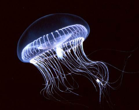
The shining protein - the north star of biochemistry. Its use today makes it possible to stain proteins and follow processes in cells that would otherwise be invisible, such as the degeneration of nerve cells in Parkinson's patients or the development of pancreatic cells in a fetus

The luminous green fluorescent protein, GFP, was first discovered in the beautiful jellyfish Aequorea Victoria in 1962. Since then, this protein has become one of the most important tools in modern use in the life sciences. With the help of GFP, the researchers were able to develop ways to observe processes that were previously invisible, such as the development of nerve cells in the brain or the spread of cancer cells.
Tens of thousands of different proteins are found in every living thing, monitoring important chemical processes in precise detail. If any protein does not function due to some mechanical reason, this usually results in disease. This is the reason why it was important for biochemists to map the role of the different proteins in the body.
This year's Nobel Prize in Chemistry was awarded to the first discoverers of the GFP, and a series of important developments that led to the widespread use of the protein as a tool for mapping the scales of life. Using DNA technology, researchers can now attach GFP to other interesting but invisible proteins. The fluorescent marker allows them to observe the movements, location and interactions of the tagged proteins.
The researchers can also follow the fate of several cells with the help of the GFP. Damage caused to nerve cells in Alzheimer's patients or how insulin-producing beta cells develop in the pancreas of a developing fetus. In one of the amazing experiments, the researchers were able to label different nerve cells in the brain of a mouse that was viewed like a kaleidoscope of colors.
The story behind the discovery of GFP was divided into three winners who had the leading roles:
Osamu Shimomurawho first isolated the GFP from the Aequorea Victoria, which drifts with the current in the western coastal area of the North American continent. He discovered that this protein glows green when illuminated with ultraviolet light.
Martin ChalfiDemonstrated the value of the GFP as an illuminating genetic tag for a selection of biological phenomena. In one of the first experiments he conducted, he illuminated six individual cells in the transparent roundworm Caenorhabditis elegans with the help of GFP.
Roger ZinnContribute to our general understanding of how the GFP lights up. It also expanded the range of colors beyond green and allowed researchers to give different proteins in a cell different colors. This allowed scientists to monitor several separate biological processes simultaneously.

One response
Good signature to all surfers and writers.