IBM scientists, who used the second most powerful computer in the world - Blue Jean, developed a method that will help in the diagnosis and treatment of bone thinning disease

Scientists at the IBM Research Laboratory in Zurich have developed a computer simulation method that will help diagnose and treat osteoporosis - the thinning of the bone (or as it is popularly called "calcium leakage"), which affects one out of three women and one out of five men over the age of 50, and is usually diagnosed after it has reached an inoperable stage to amend.
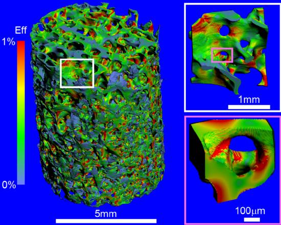
Using IBM's Blue Gene supercomputer, IBM scientists and researchers at the Swiss Federal Institute of Technology (ETH) were able to present the most detailed simulation ever performed of bone structure in the human body, down to the level of the internal microstructure of each segment Closed. This high level of separation opens the way for research in the field of the strength of these bones, which was not possible before. The unique technological achievement may pave the way for the development of better tools for the diagnosis and treatment of osteoporosis.
Diagnosing osteoporosis as early as possible is essential in order to prevent the continuation of the disease process. The groundbreaking simulation may significantly improve the ability of doctors to better treat fractures, and to analyze the level of bone fragility and strength at each stage of the disease. Thus, it is possible to decide which tools should be used to treat each patient, in order to prevent fractures even before they occur.
Osteoporosis is the most common bone disease in the world. The disease affects 75 million people in the US, Europe and Japan, and many tens of millions more in the rest of the world. The costs arising from the disease, in terms of the price of treatments, hospitalizations and burden on the health system, constitute the second largest section of the medical budget, after the costs of cardiovascular diseases.
The disease is characterized by a decrease in bone density, and leads to a high risk of fractures. It causes pain, limits the movement and activity of older people. In many cases, the disease is not diagnosed until the first fracture occurs - but then it is already in an advanced stage, and requires the use of implants or expensive surgical procedures in order to treat these fractures or prevent the next fracture.
Osteoporosis is currently diagnosed by measuring bone density, using special X-rays or computerized scanning (CT) technologies. However, studies state that the study of bone mass with the tools available today only shows a moderate level of accuracy in answering the real question - what is the strength of the measured bone. This inaccuracy is due to the fact that bones are not a solid and completely complete structure: inside the hard and complete outer shell, the bones are characterized by a spongy internal structure. This microstructure is responsible for the bone's ability to carry heavy loads - and its measurement provides the best indication of the bone's true strength.
In order to develop an accurate and fast method for automatic analysis of bone strength, they teamed up experts in the field of mechanical engineering and process engineering at the Swiss National Institute of Technology in Zurich together with supercomputer experts at the IBM research laboratories operating in the city. The groundbreaking method developed by the joint team combines bone density measurement with large-scale mechanical analysis of its internal microstructure.
With the help of a large-scale simulation, and massive parallel processing, the IBM scientists manage to present a "map of load zones", and analyze the changes that apply to the loads the bone is under. This map gives doctors an exact answer to the question of where in the bone the fracture may occur - and what is the expected load to cause such a fracture. On the basis of the detailed and more accurate analysis than ever before, the doctor can determine and adapt a surgical process, implant plates to support the bone and place the implant at the most correct point.
IBM's Blue Gene /L supercomputer allowed the research team to perform the first simulation of a real human bone sample, measuring 5x5 mm. 20 minutes of processing time were required for the computer to generate information in the volume of 90 gigabytes, which provides a description and analysis of the piece of bone at the most detailed structural level.
IBM predicts that within ten years the capabilities that characterize the current generation of supercomputers will be available at the level of desktop systems, and detailed simulations of bone strength will become a daily concern in the computed tomography process.
In order to complete the processing, advanced mathematical algorithms were required that allow solving problems at the highest speed. The present work is a first basic step on the way to clinical use in large-scale bone simulation. Now, the scientists plan to expand their computational technologies beyond the analysis of the static strength of the bone, in order to perform a simulation at the level of the individual patient.

2 תגובות
What does IBM get out of all this? Are they doing it for charity?
The bone was described using a surface unit instead of volume.