The discovery may contribute to the development of treatments for deafness that will be based on the regeneration of hair cells in the ear * According to the researchers, this is a change of perception in developmental biology: the researchers found that the development of the array of hair cells in the inner ear is similar to the organization of atoms that form a crystal - a known process in physics, which has not yet been identified in biology

A study led by Prof. David Sheprincek, an expert in developmental biology from the School of Neurobiology, Biochemistry and Biophysics in the George S. Wise Faculty of Life Sciences of Tel Aviv University, revealed for the first time a physical mechanism involved in the development of the inner ear in mammalian embryos.
Prof. Sheprincek: "This is the first time that a biological development process has been discovered that is driven by mechanical forces and is very similar to a known physical process: the organization of hair cells in the inner ear is similar to the organization of atoms that form a crystal. This is a revolutionary finding that changes fundamental concepts in the world of developmental biology."
Participating in the multidisciplinary research were: Roy Cohen and Liat Amir-Zilberstein from the laboratory of Prof. Sheprincek, Prof. Keren Avraham from the Sackler School of Medicine and other researchers from the Faculty of Exact Sciences and the Segol School of Neuroscience at Tel Aviv University. Researchers from Switzerland and Japan also participated. The article was published in the prestigious journal Nature Communications in October 2020.

Hairs act as sensors for the sound waves
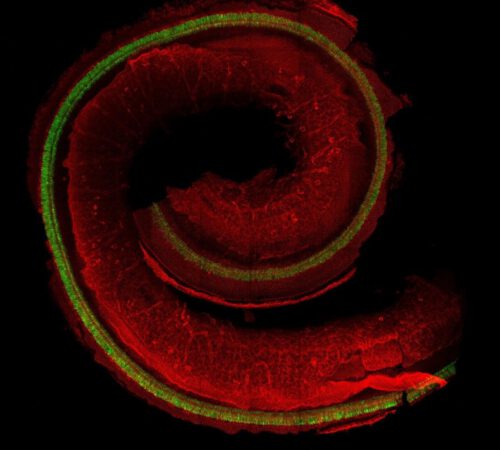
Prof. Shaprincek explains: "The ear of a mammal consists of three parts: the outer, middle and inner ear. Inside the inner ear is the cochlea - a spiral structure along which hair cells are embedded - cells on which tiny hairs act as sensors for the sound waves reaching the ear. In response to the sound waves hairs oscillate and transmit electrical signals to the brain, and this is how we hear. The hair cells are arranged along the cochlea in a unique way: four arranged rows of alternating hair cells and supporting cells. This strict order is important because different areas along the cochlea are responsible for detecting different sounds. Such an organized array is unusual in the body, and in fact it is the most organized tissue in the body of a mammal. We were looking for the mechanism that causes hair cells to arrange themselves in this way during embryonic development. To this end, we conducted a multidisciplinary study that combined two innovative developments: a new imaging technology and a simulation using a mathematical-physical model."
To monitor the development of the hair cell array in the embryo, the researchers examined mouse embryos at different stages of pregnancy. In the youngest embryos, an unordered and unsorted layer of cells was found - primary cells whose function has not yet been defined. Gradually, the cells begin to differentiate into hair cells and supporting cells, and later they organize, until the final and orderly structure of the hair cells in the cochlea is formed.
Prof. Shaprincek: "Until today, most studies on the development of hair cells have focused on the differentiation processes of the cells, which are controlled by intercellular communication. We believed that this was not enough, and asked to examine the stage after differentiation: how the cells organize themselves in space and create an orderly structure." For this purpose, the researchers developed a new imaging technology that relies on observation with a unique microscope, and enables continuous monitoring in 24D, XNUMX hours a day, of tissue development - a process that lasts a few days in mouse embryos. The unique imaging method made it possible to track the development of the inner ear and create videos of the process.
The differentiation process in the fetal ear
Prof. Sheprincek: "This is the first time that the process has been observed continuously and with high resolution. We observed tissue that had already differentiated into hair cells and supporting cells, but still remained 'messy' - that is, it had not yet organized into an orderly array. Using our method, we saw that a physical process known as a 'shearing process' takes place in the tissue: neighboring cells, called 'Hansen cells', move in one direction, and exert shearing forces on the hair cells - mechanical forces that work parallel to the cell layer. This process compresses the tissue, causes dynamic exchanges between hair cells and supporting cells, and ultimately leads to the cells being organized in an orderly array.
In the next step, the researchers relied on their findings to create a computer simulation - a mathematical-physical model of the process. The model revealed that two main mechanical forces act on the hair cells during the organization phase: shearing forces, which cause compression and movement of the hair cells within the tissue, and repulsive forces between the hair cells, which prevent them from getting too close to each other. Prof. Sheprincek: "We were surprised to discover that the process of organizing the hair cells in the cochlea is very similar to a well-known physical process: the organization of atoms during the formation of a crystal. Just as atoms form an ordered crystal when external forces are applied to them, so the hair cells and the supporting cells organize themselves into an ordered pattern in response to mechanical forces applied to them. This is a completely new concept in the field of developmental biology. The insights that emerge from our research illuminate a new direction for future studies, also regarding the development processes of other organs."
Prof. Ronan Peretz, Director of the Otology Unit and the Cochlear Transplant Center at the Shaare Zedek Medical Center affiliated to the Faculty of Medicine of the Hebrew University who was not involved in the research, notes that the research findings may also have medical implications: "Hair cells are created in the fetus, and do not regenerate during life. In many cases, hearing loss and even deafness result from the death of hair cells in the inner ear. In recent years, many efforts have been made to develop treatments with the help of genetic therapy that will encourage the regeneration of hair cells, with the aim of improving the hearing ability of deaf people. The research may make an important contribution to understanding the process towards the creation of this renewal."
More of the topic in Hayadan:

2 תגובות
Well done David and the team, it's exciting. We hope that thanks to this discovery, one day there will be no deaf people, like they managed to help AIDS patients, and maybe one day they will be able to restore myelin to MS patients
Your work is sacred work
Thanks
Respect to researchers