Tears do not flow by themselves - Weizmann Institute scientists reveal the surprising mechanism by which saliva, tears and digestive juices are secreted
When we see an appetizing dessert, bite into an apple or burst into tears, cells in our body secrete special fluids: saliva, digestive juices or tears. The ways in which these fluids and other substances are secreted from the body's cells have been studied for almost a century and are described in detail in textbooks. Still, Dr Uri Avinaam and Prof. Ben-Zion Shiloh The Weizmann Institute of Science believed that the classical descriptions were lacking.

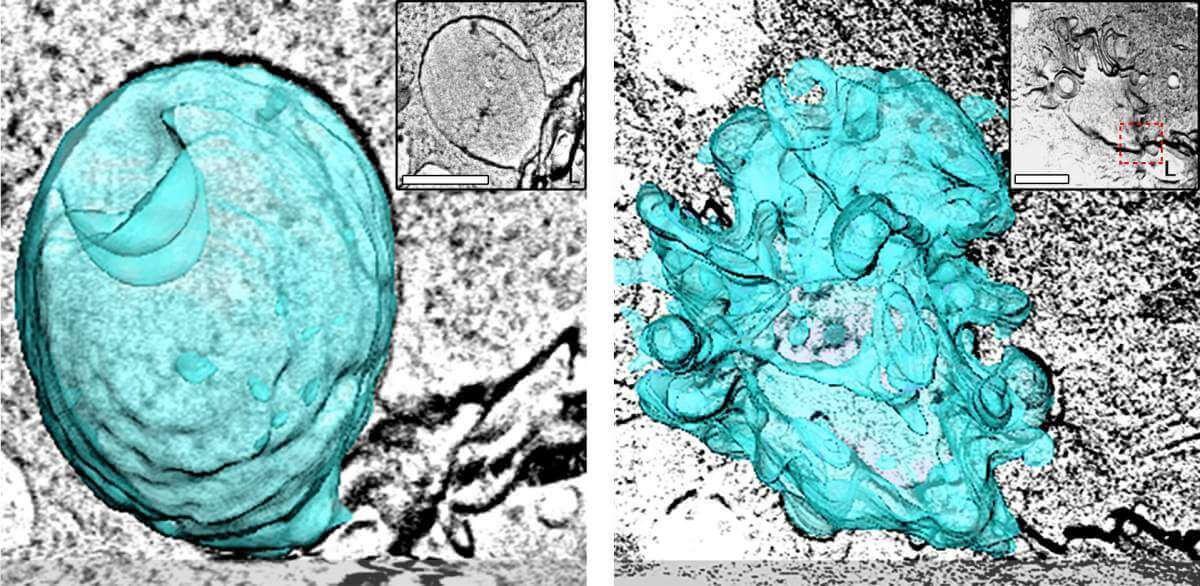
When a cell secretes substances, whether it's waste, hormones or neurotransmitters, it does so using tiny bubbles called vesicles. These vesicles fuse with the cell membrane in the following way: their membranous envelope becomes part of the cell membrane, while their contents spill out of the cell. This classic description does faithfully describe the secretion mechanism in which tiny bubbles with a diameter of less than 100 nanometers (billionths of a meter) are involved, but cells called "professional secretors" - for example those that secrete digestive enzymes, moisturizers or lubricants - pack their cargo in bubbles 100 times larger , whose diameter reaches 10 microns (millionths of a meter). The secretion from these bubbles is much slower and lasts for minutes, as opposed to thousandths of a second in the case of the tiny bubbles. These "giant" bubbles are indeed much more economical and efficient than their tiny sisters when it comes to excreting a substance in large quantities, but do they excrete their contents using the same familiar mechanism?
Prof. Shiloh's laboratory in the Department of Molecular Genetics studies the salivary glands of fruit fly larvae as a model for the secretion of substances from large vesicles. Dr. Avinaam's laboratory from the Department of Biomolecular Sciences investigates cell membrane remodeling mechanisms, using advanced imaging technologies. In joint conversations, the two scientists hypothesized that the well-known and well-known secretion mechanism probably does not fit the description of the secretion from large vesicles, since if the envelope of these vesicles merged with the cell membrane, the cell would swell and deform, and at some point even stop functioning.
To solve the mystery, Prof. Sheila and Dr. Avinaam teamed up with Dr. Kamalesh Komari, a post-doctoral researcher in both laboratories, who led the research. The scientists used XNUMXD electron microscope imaging combined with other imaging methods to examine secretory cells in both the salivary glands of the fruit fly larvae and the mouse pancreas. Nadav Sher, Tom Biton and Dr. Eil Schechter participated in the study.
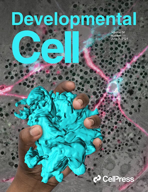
"We discovered that large bubbles, unlike the tiny ones, secrete substances through a mechanism that was not known until now - a completely different mechanism from the one described in the textbooks," says Dr. Avinaam. In fact, the researchers discovered that when large bubbles adhere to the cell membrane, they do not merge with it at all, but rather secrete their contents in a different way - similar to the way an inflated sea ball is deflated and then compressed back into a bag; In English, the scientists attached the word "crumpling" to the mechanism - crushing or crumpling. Their experiments revealed that a network of proteins called actomyosin wraps the bubble, crushes, crumples and squeezes its contents out of the cell through a narrow opening - all while maintaining the bubble's separation from the cell membrane. Cellular molecular machinery is then sent to the site to produce thousands of small bubbles from the crumpled shell in order to recycle its components.

"These findings open up new directions of research in regards to secretions from the cell that are carried out using large bubbles, with an emphasis on mechanisms that maintain the integrity of the cell membrane during the secretion", says Prof. Sheila. Since the new mechanism was discovered both in fruit fly larvae and in mammals, the scientists speculate that it also exists in humans and may be involved in the development of various diseases. Dr. Kamalesh explains: "It is very possible that defects in the crushing mechanism of the bubble are involved in diseases in which there is a lack of secretions, for example dry eye syndrome or when there are excess secretions, such as cystic fibrosis. Such defects may also play a role in diseases that affect internal surfaces of the body that require constant lubrication, such as the internal lining of the digestive system."
in bright colors
Already in high school in Haifa, Dr. Uri Avinaam crossed the lines and moved from the student's chair to behind the teacher's desk. It happened when the biology teacher fell ill during matriculation preparations, and the students asked Uri to replace her; Needless to say, the teacher did not object. "It was always very natural for me to study and teach biology," he says.
While studying for a bachelor's degree in biology and biochemistry at the Technion, he was fascinated by cell membranes and other biological membranes that are essential to our existence, and was looking for a summer internship that would help him understand the interrelationships between membranes and the proteins embedded in them. In the laboratory he finally joined, he was amazed to see for the first time cells glowing with phosphorescent colors using fluorescence - then a relatively new method in biology. "This sight is etched in my memory," he explains.
While studying in a direct path to a doctorate at the Technion, and then in post-doctoral studies at the European Laboratory for Molecular Biology in Heidelberg, Germany, Dr. Avinaam found a way to combine the magic of the two fields: he began to study membranes using advanced imaging methods.
In his laboratory at the Weizmann Institute of Science, Dr. Avinaam studies cell membranes using fluorescence, as well as electron microscopy and other advanced methods. It focuses on the processes related to cell fusion and the transport of molecular charges through membranes, as they are manifested, for example, in the fusion between a sperm cell and an egg, in the development of skeletal muscles or in the secretion of substances from the cell through membrane-encased vesicles.
Dr. Avinaam lives on the institute's campus with his partner Yuval, an aerospace engineer, and with their three-year-old twins - a boy and a girl.
More On the subject of the science website:
