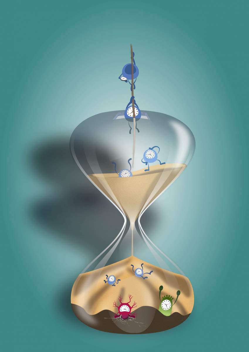
The wonderful world where the first embryonic cells are formed is now more accessible to us than ever before
Each of the millions of cells that make up our body and that of other animals, knows its place and function very well and integrates precisely in the development and operation of the entire organic body. Scientists follow this fascinating dynamic with great interest, when it functions perfectly or when it is impaired by diseases, such as cancer, and aging.
A particularly suitable time point for the study of these complex processes is at the beginning of the development of an embryo, when new types of cells emerge at an incredible speed. Groups of individual cells adapt themselves to diverse roles, and in the process reproduce into hundreds, thousands and finally millions of cells - which make up the initial structure from which all the organs of the embryo will develop (a process known as gastrulation).
Normally, cells form and change relatively slowly, subject to many environmental influences. The first moments of the fetus are characterized by development processes that repeat themselves precisely, in a kind of orchestrated dance of millions of participants. Observing these processes makes it possible to search for answers to the big questions of the world of cells and to learn how and when they adopt different roles and become part of the brain, bone or digestive system. However, because the fetus is a tiny structure that grows rapidly and is protected by the mother's body, scientists have usually had to study it in indirect ways.
Dr. Yoav Misher and researchers from the laboratory of Dr Yonatan Stelzer from the Department of Molecular Biology of the Cell at the Weizmann Institute of Science, together with Dr. Markus Mittenzweig and researchers from the group of Prof. Loaded condition From the Department of Computer Science and Applied Mathematics and from the Department of Biological Control, find new way follow the complex process. They were helped by technological breakthroughs from recent years, which allow mapping of genetic activity patterns at the single cell level. Researchers around the world have already created detailed atlases of all types of active cells in various animals, but the researchers at the Weizmann Institute estimated that a deeper understanding of embryonic development requires drawing these atlases with reference to the time dimension.
Because genetic mapping at the single cell level impairs the continued normal functioning of the cell, researchers were previously forced to make educated guesses about the development process of the cells. For example, they assumed that the cells take the shortest path from the base state to the sorted state. "If fetal development is a dance," says Dr. Mittenzweig, "then in previous models we were forced to assume that every dancer is a soloist who is looking for the shortest way from entering and exiting the stage." However, adds Dr. Misher, "this is not compatible with developmental biology as we understand it based on years of experiments."

In the current study, a unique combination of information was created from tens of thousands of individual cells of mouse embryos, together with pictures and physical measurements of the whole embryos. In this way, the developmental process can be reconstructed through the identification of tiny changes in the shape of the embryo, as has been the practice in this research for decades, in combination with a mathematical model that takes into account the genetic activity of the cells in each individual embryo. The result is a continuous "film" that describes the joint transformation of the cells of the embryo and which consists of a collection of discrete "cellular photographs".
The researchers noticed that while some of the cells run straight to the goal, for example creating vital organs for the fetus, some of the cells retain extensive developmental potential. The study confirms the hypothesis that cells develop in a process of continuous change, and that there is a great potential for change not only in their first moments, but also in later stages. This potential exists within the framework of a complex structure, with many and changing mutual effects between the cells, along with a change in the role of each gene depending on the time and the cell in which it is expressed.
Figuratively, it can be said that the resulting model describes how the cells move in a kind of virtuoso dance in which each dancer, that is, each cell, does not move in a predetermined route - but connects to very dynamic ensembles with the thousands and millions of other cells around it. The tissues formed in this way arrange themselves in a complete organic structure and in a magical harmony, which repeats itself over and over again every time a new embryo is formed.
The research presents a new and sophisticated work tool with the help of which it will be possible to continue to follow the world of the cells and to understand why and how they assume different roles, how they affect their environment and how it affects them, and why sometimes their activity goes wrong in a way that harms the functioning and existence of the living being.
