Weizmann Institute scientists go on research trips following the production processes of composite materials in the living world

Humans are different from snails and molluscs, but it is not certain that this difference is expressed only in advantages. A little modesty would have been helpful here. Certain snails can, for example, complete and regenerate damaged nerve fibers. If humans were able to do this, a cure would be found for many diseases caused by nerve damage or nerve cell degeneration. But that's not the only thing worth learning from shellfish. Another striking example is the incredibly beautiful and strong structures (shells and clams) that certain molluscs make. These crystalline structures, and their construction processes, are an inspiration to scientists who develop composite materials that are extremely light and strong, used, among other things, for medical purposes, such as artificial orthopedic implants.
The importance of composite materials has led many scientists to research trips following the production processes of these materials in the living world.
These studies focused both on invertebrate animals, such as molluscs, and on the bones of vertebrates. Recently, a connecting line was discovered between the two types of creatures being studied: it turns out that certain methods of operation that molluscs use to build shells are also used by vertebrate animals, including humans.
The bone does not grow in one direction; It develops layer upon layer, and its structure undergoes continuous changes - processes that make it difficult to distinguish between new material and old material. Scientists who wish to follow the process of bone development encounter additional difficulties, arising from the position of the bone within the living body. It is known that in the process of bone development, which is a composite material, the crystalline mineral material is placed inside a "casting mold" made of protein fibers; But how these crystals form is controversial. The simplest and most obvious explanation was that the minerals settle directly out of solution, similar to how stalagmites and stalactites are formed. This was also the accepted hypothesis as to the way in which shells are formed, but findings published in the late 60s raised the first hints that the solution lies elsewhere.
The answer began to become clear in studies done on molluscs, which showed that the process of building their shell includes an intermediate stage, in which the animal produces amorphous mineral particles (that is, disordered, as opposed to a crystal that has an ordered structure). These particles are transported from the body of the mollusc to the place where the shell is built, and undergo a structural change there that turns them into a strong crystal. Research by Prof. Leah Eddy and Prof. Steve Weiner, from the Department of Structural Biology at the Weizmann Institute of Science (who examined the mechanisms of the formation of spines in sea urchins), and by other scientists from different parts of the world, showed that this is a general phenomenon, which exists in rigid structures in invertebrate animals more Prof. Eddi: "The original hypothesis, of the deposition of mineral material from a solution, required the flow of huge amounts of solution outside the body of the mollusk. The process we identified, in which the 'bricks' and 'cement' are transported in their solid form to the construction site, is much more efficient and logical - in terms of the animal's energy economy."
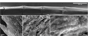
But the question regarding the ways of formation of bones in vertebrate animals remains. The attempts to identify an amorphous mineral substance, whose life span is limited, inside a bone that is in stages of growth, did not go well, and in any case failed to provide unequivocal evidence of the correctness of the theory. Research student Yulia Mahamid, Prof. Lea Addi and Prof. Steve Weiner, from the Department of Structural Biology at the Weizmann Institute of Science, recently succeeded in finding a special biological system that allows to follow, step by step, the various stages of the bone formation process. The scientists chose to study the fin bone of a zebrafish, which grows "from the inside out" throughout the fish's life. The fin is made like a "bone fan", each of which consists of several sections - so the section at the end of the fan is the newest. Because of this, the chapters can be considered as a "timeline" showing different stages in the development of the bone. In addition to this, the low temperature in which the fish lives slows down the development of the mineral substance in the bone, and makes it easier to monitor it.
Through a combination of scanning and penetrating electron microscopy, and other advanced methods, the scientists were able to definitely identify spherical particles, made of amorphous mineral material, and distinguish it from the crystalline mineral material. The findings, recently published in the scientific journal "Records of the National Academy of Sciences" of the USA (PNAS), show that in the "young" chapters
of the bone, the amorphous material constitutes about half of the total mineral material, and that its amount is getting smaller as the bone matures - at the same time as the general amount of the crystalline material increases. Crossing data obtained from the two microscopes made it possible to track certain locations on the bone, and to indicate the possibility that amorphous particles turn, over time, into crystals.
These findings shed light on a long-standing mystery, and in a broader aspect, they provide new information and a unique point of view on basic biological processes in which the mineral substances that make up the hard tissues, such as bones and teeth, are formed. These processes occur in all vertebrate animals, including humans. The scientists hope that a thorough understanding of these processes will help in the future to promote the healing options of genetic diseases that cause abnormal bone construction.

Above: Scanning electron microscope photograph of one "finger" from the fan that makes up the skeleton of the zebrafish's fin. Below: different stages of bone development. In the earliest stage of development (right) the bone is composed mainly of spherical amorphous mineral particles. In the next step (in the center) you can see round particles (marked with white arrowheads), and next to them crystals (black arrows). In the adult stage (left) the bone consists mainly of crystals
