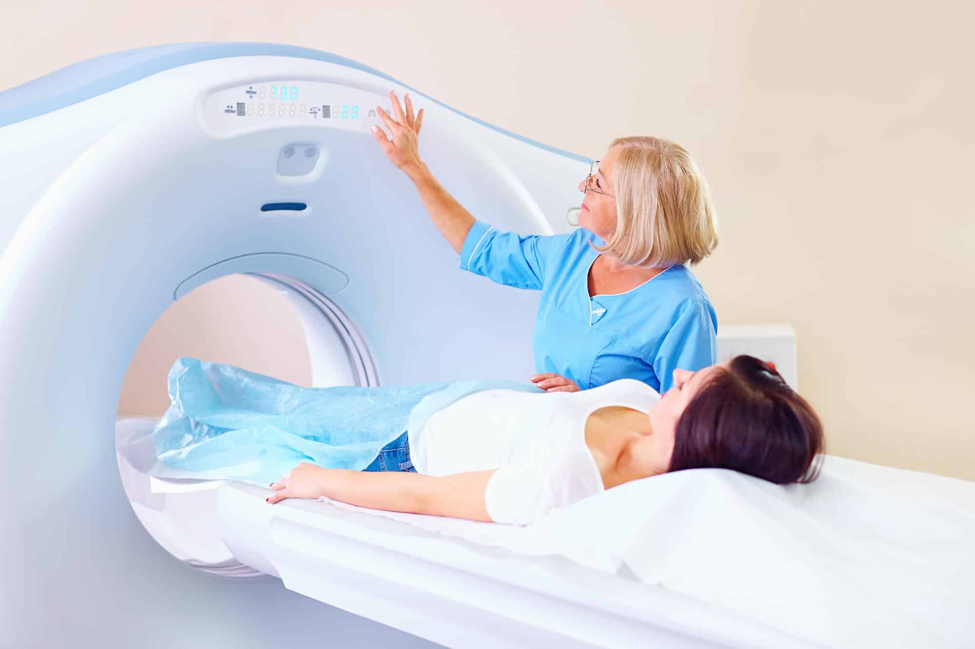
As the years go by, new types of sequences and protocols are developed and are joined by improvements in software and hardware - for example, you can see the great development in recent years in the field of cardiovascular imaging, bowel imaging and more

The diagnosis with the help of an MRI scan is a fertile ground for research. As the years go by, new types of sequences and protocols are invented and to them are added the improvements in software and hardware - for example, you can see the great development in recent years in the field of cardiovascular imaging, intestinal imaging and more. As of after the middle of 2022, new MRI scans, sequences, and protocols have been added, allowing special scans to be performed with better quality than before. We will present a selection of them in this article and in other articles below:
MRI Sialography- MRI scan to demonstrate salivary glands
MRI scan sialography (Sialography) is a scan that examines the salivary glands - especially the parotid glands, which are located in front of the ear. It can be a complementary test to other tests performed on the salivary glands such as ultrasound, X-ray sialography, sialography under reflection with a suitable contrast material and CT sialography. Even in the past it was of course possible to perform an MRI scan of the salivary gland area, but the invention of new types of sequences have made the test today a fairly sensitive and reliable test for evaluating the salivary glands. During the test, we use sequences with T2 enhancement (such as RARE, CISS, FISP - these sequences are also used in MRCP and MR Urography scans). These sequences clarify the fluid in the gland and demonstrate the ducts well, without the need for the injection of contrast material (although the injection of contrast material only improves the image).
The indications for performing an MRI sialography scan are, for example, in the case of sialolithiasis (Sialolithiasis - a chronic disease in which a stone forms in the salivary duct and causes repeated inflammation of the salivary glands), obstruction of the salivary duct, in identifying a cause for sialadenitis (Sialadenitis - inflammation of the salivary glands that causes the narrowing of the salivary ducts) and in suspicion Sialoangiectasis - expansion of the salivary ducts, which is caused by infection or destruction of the salivary gland following chronic inflammations and autoimmune diseases.
Advantages MRI Sialography They are a relatively fast acquisition of the image, a non-invasive procedure, possible assessment of other glands in the area, good spatial resolution and a comfortable position of the subject. Also, there is no exposure to X-ray radiation and there is no need to use a tube as is done in conventional sialography or under mirroring.
Disadvantages MRI Sialography By and large, they are general contraindications for performing an MRI such as pacemakers, implants, a test that can cause claustrophobia, and the presence of dental fillings, implants and bridges that can impair the quality of the test. Also, in this scan it is possible to demonstrate only first and second order branches of the salivary ducts.
- You can read more about the MRI sialography scan In this link
MRN - MR Neurography - nerve imaging test
This test was originally intended to be used for imaging peripheral nerves (peripheral nerves). She evaluates peripheral nerve disorders and also locates and grades nerve injuries. The visual information of the exact location and extent of nerve abnormalities provided by MRN makes it a powerful tool that helps doctors and surgeons reach an accurate diagnosis and decide on further medical or surgical treatment. Sequence improvements in recent years have resulted in the ability to perform this test much more reliably than before.
The test can be performed anywhere there are peripheral nerves such as the shoulder, neck and chest area (brachial plexus), sacral plexus (is part of the larger lumbosacral plexus, supplies motor and sensory nerves to the hind thigh, most of the lower leg, the entire foot and part of the pelvis) , wrist nerves, ankle nerves and more. The MRN test is also good for demonstrating the cranial nerves (the 12 cranial nerves) - for example for demonstrating the trigeminal nerve (Nervus trigeminus) and is also possible for other uses.
One of the types of protocols used in this test is the TrueFISP scanner as it is called at Siemens (or Fiesta at GE and balancedFFE at Philips).
- You can read more about this test In this link On the Siemens website
Glutamate Chemical Exchange Saturation Transfer (GluCEST) MRI - scan to diagnose encephalitis (inflammation of the brain)
Encephalitis is a common inflammatory disease of the central nervous system that endangers human health due to the lack of effective diagnostic methods, which leads to a high rate of misdiagnosis and mortality. Glutamic acid (also known as glutamate; one of the 20 amino acids common in nature) is closely involved in the activation of cells called microglia (Microglia - a type of glial cells, non-neuronal support cells for neurons) and the activation of these cells serves as a key player in brain inflammation.
An MRI scan called GluCEST (glutamate chemical exchange saturation transfer) can be used for early diagnosis of encephalitis by detecting the concentration of glutamate. Inflammatory areas of the brain demonstrated a particularly intense GluCEST signal due to the presence of high concentrations of glutamate there, while after treatment with intravenous immunoglobulins, this signal decreased following an improvement in the state of inflammation.
In conclusion, glutamate plays a role in encephalitis, and the GluCEST test signal has the potential to be an in vivo imaging biomarker for the early diagnosis of encephalitis.
- You can read more about this test In this article
Over the years, many other interesting techniques and protocols have been created that improve the imaging and diagnostic ability. For example, we can mention the MR Elastography scan, a non-invasive technique designed to assess the stiffness of soft tissues. This technique can be performed in any MRI scan, such as a liver MRI scan in order to define the level of fibrosis in it. It is also possible to mention the great development in the field of diffusion sequences in MRI, which today enable the detection of pathologies in a much more precise way than before in many organs of the body.
In conclusion, there have been many developments in the field of MRI in recent years and the research towards further developments and improvements in the fields of hardware, software, protocols and imaging is constantly taking place in order to continue one of the biggest revolutions in the field of medical imaging and research in medicine. We will try to keep you updated on the many developments in the field in the future as well.
The author of the article: Ofer Ben Horin, who has about 20 years of experience in applications, drug research and training in the field of MRI. Author of the bookMRI the complete guide-medicine and physics meetOn the website www.mriguide.co.il
More of the topic in Hayadan:
